5UIG image
Deposition Date 2017-01-13
Release Date 2017-02-08
Last Version Date 2024-11-06
Entry Detail
PDB ID:
5UIG
Title:
Crystal structure of adenosine A2A receptor bound to a novel triazole-carboximidamide antagonist
Biological Source:
Source Organism:
Homo sapiens (Taxon ID: 9606)
Escherichia coli (Taxon ID: 562)
Host Organism:
Method Details:
Experimental Method:
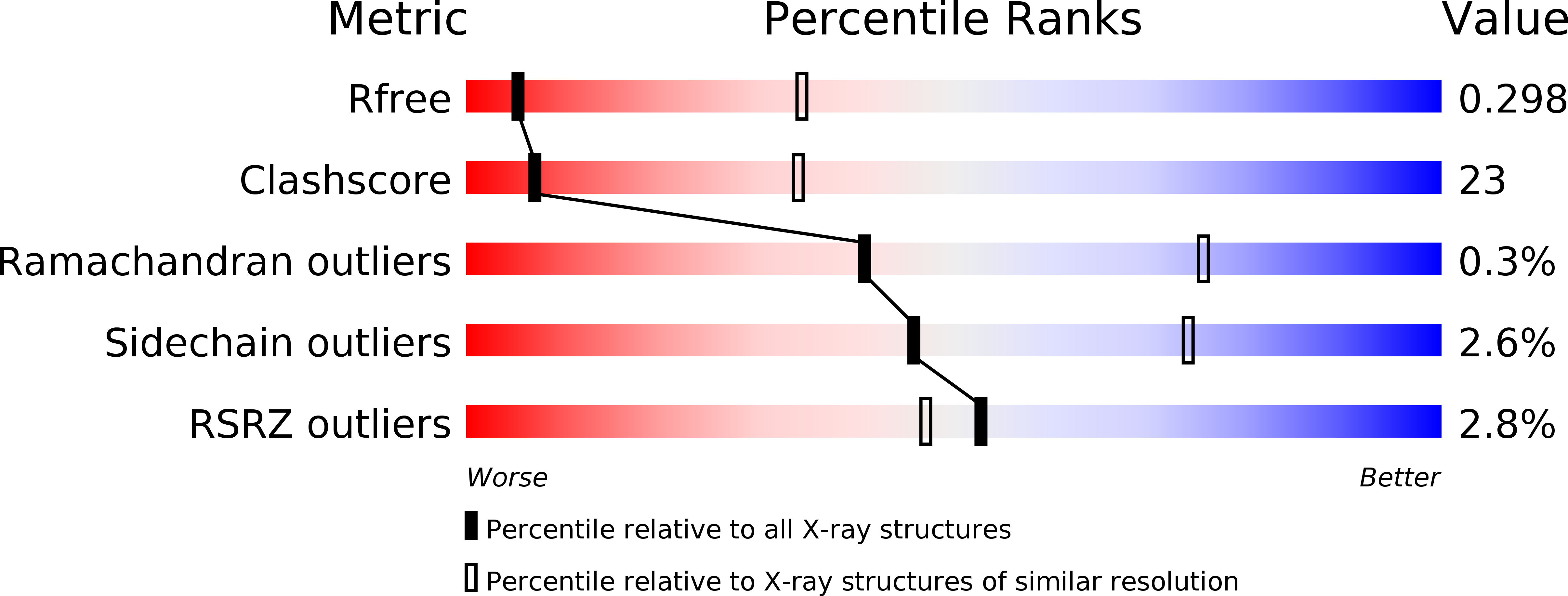
Resolution:
3.50 Å
R-Value Free:
0.29
R-Value Work:
0.27
R-Value Observed:
0.27
Space Group:
P 41 21 2
Macromolecular Entities
Protein Blast
Polymer Type:polypeptide(L)
Molecule:Adenosine receptor A2a,Soluble cytochrome b562,Adenosine receptor A2a
Gene (Uniprot):cybC, ADORA2A
Mutagens:M1007W, H1102I, R1106L,M1007W, H1102I, R1106L,M1007W, H1102I, R1106L
Chain IDs:A
Chain Length:444
Number of Molecules:1
Biological Source:Homo sapiens, Escherichia coli
Primary Citation
Crystal structure of the adenosine A2A receptor bound to an antagonist reveals a potential allosteric pocket.
Proc. Natl. Acad. Sci. U.S.A. 114 2066 2071 (2017)
PMID: 28167788 DOI: 10.1073/pnas.1621423114

Abstact

The adenosine A2A receptor (A2AR) has long been implicated in cardiovascular disorders. As more selective A2AR ligands are being identified, its roles in other disorders, such as Parkinson's disease, are starting to emerge, and A2AR antagonists are important drug candidates for nondopaminergic anti-Parkinson treatment. Here we report the crystal structure of A2A receptor bound to compound 1 (Cmpd-1), a novel A2AR/N-methyl d-aspartate receptor subtype 2B (NR2B) dual antagonist and potential anti-Parkinson candidate compound, at 3.5 Å resolution. The A2A receptor with a cytochrome b562-RIL (BRIL) fusion (A2AR-BRIL) in the intracellular loop 3 (ICL3) was crystallized in detergent micelles using vapor-phase diffusion. Whereas A2AR-BRIL bound to the antagonist ZM241385 has previously been crystallized in lipidic cubic phase (LCP), structural differences in the Cmpd-1-bound A2AR-BRIL prevented formation of the lattice observed with the ZM241385-bound receptor. The crystals grew with a type II crystal lattice in contrast to the typical type I packing seen from membrane protein structures crystallized in LCP. Cmpd-1 binds in a position that overlaps with the native ligand adenosine, but its methoxyphenyl group extends to an exosite not previously observed in other A2AR structures. Structural analysis revealed that Cmpd-1 binding results in the unique conformations of two tyrosine residues, Tyr91.35 and Tyr2717.36, which are critical for the formation of the exosite. The structure reveals insights into antagonist binding that are not observed in other A2AR structures, highlighting flexibility in the binding pocket that may facilitate the development of A2AR-selective compounds for the treatment of Parkinson's disease.

Legend

Protein

Chemical

Disease

Primary Citation of related structures