Deposition Date
2017-01-04
Release Date
2017-05-10
Last Version Date
2025-04-02
Entry Detail
PDB ID:
5UFK
Keywords:
Title:
Structure of the effector protein SidK (lpg0968) from Legionella pneumophila
Biological Source:
Source Organism(s):
Legionella pneumophila (Taxon ID: 446)
Expression System(s):
Method Details:
Experimental Method:
Resolution:
2.30 Å
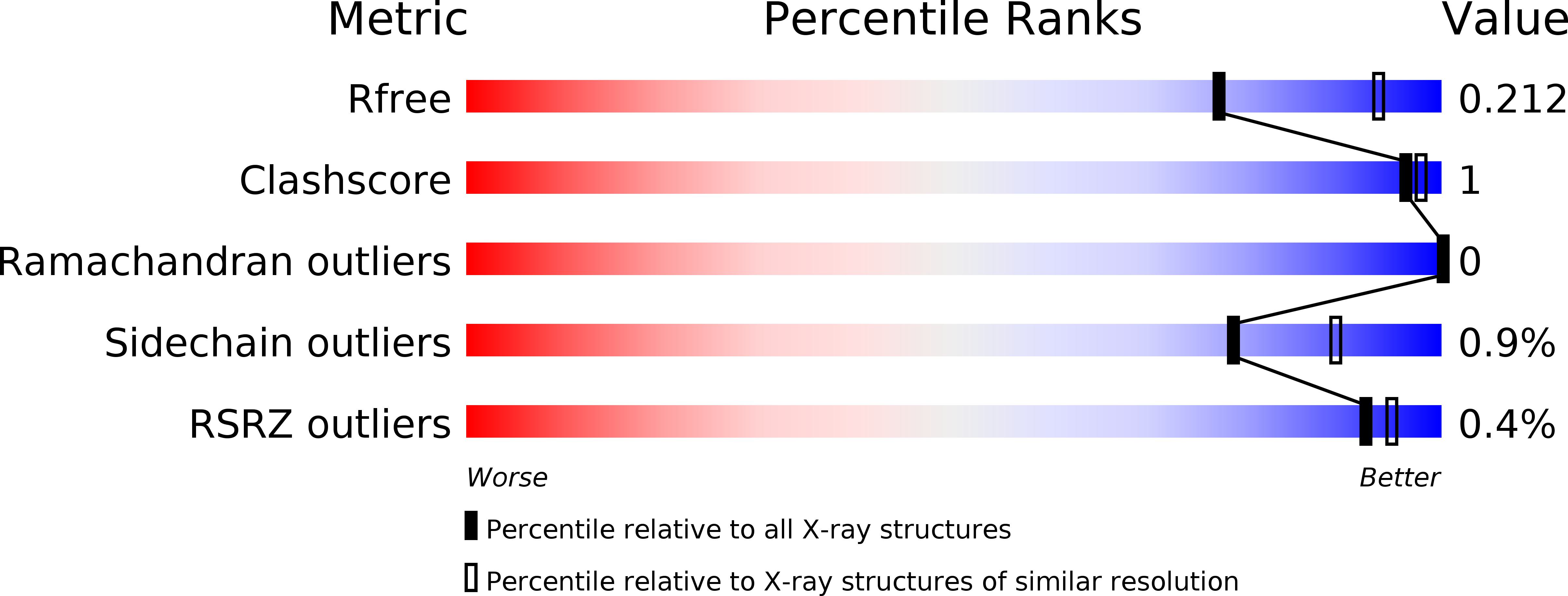
R-Value Free:
0.21
R-Value Work:
0.17
R-Value Observed:
0.17
Space Group:
P 43