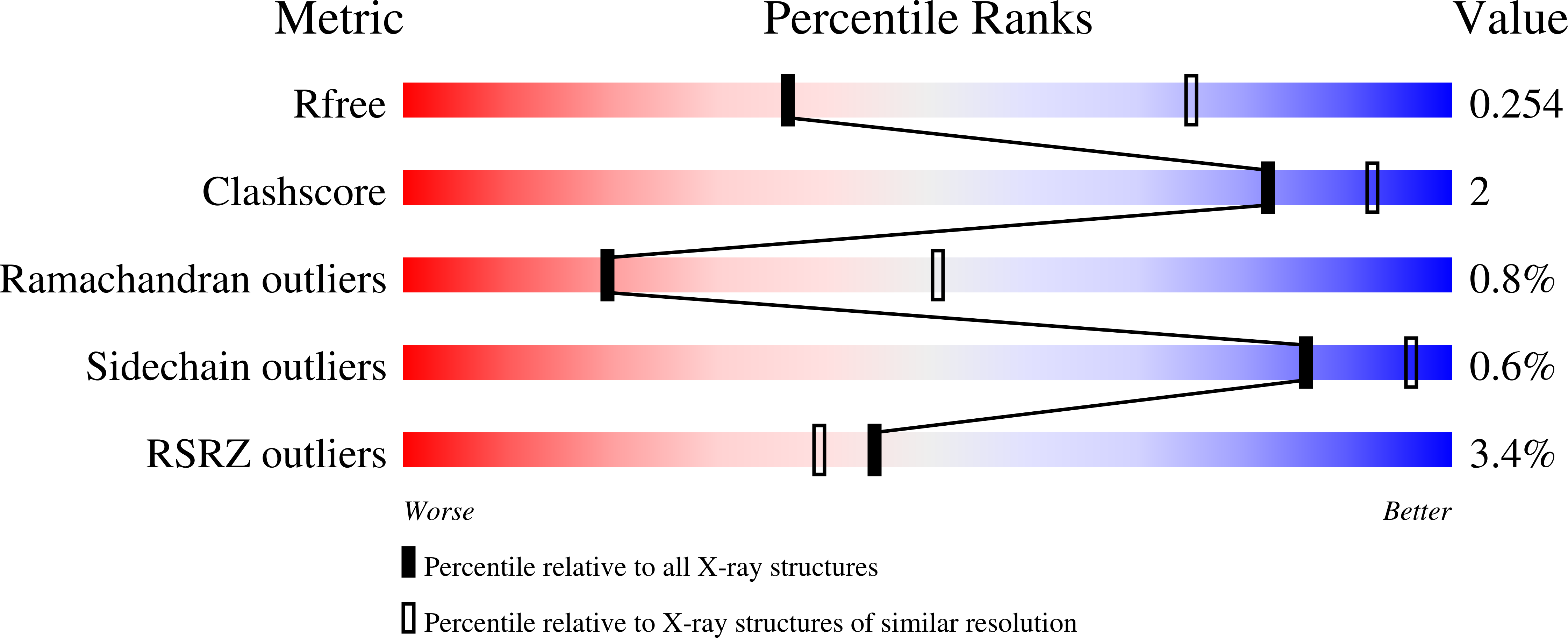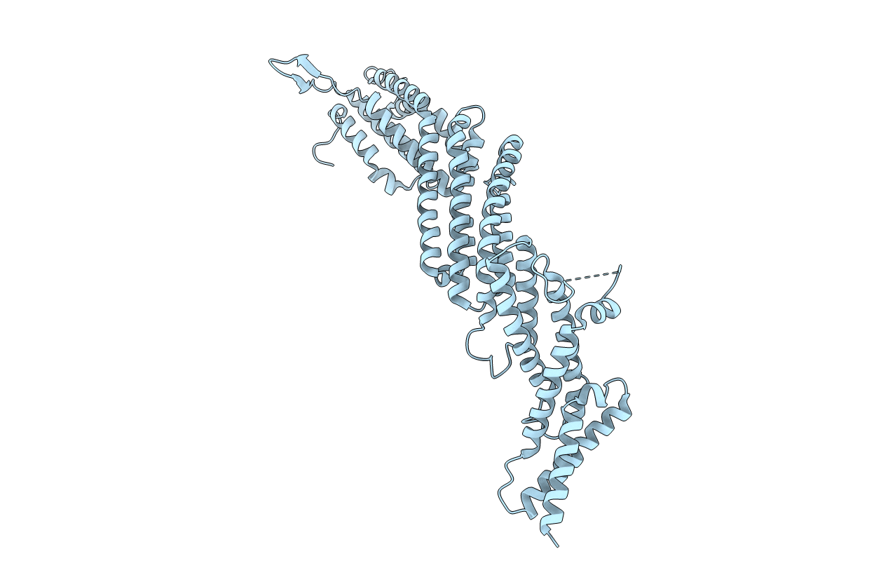
5UF7 image
Entry Detail
PDB ID:
5UF7
Keywords:
Title:
CRYSTAL STRUCTURE OF MUNC13-1 MUN DOMAIN
Biological Source:
Source Organism:
Host Organism:
PDB Version:
Deposition Date:
2017-01-03
Release Date:
2017-02-15
Method Details:
Experimental Method:
Resolution:
2.90 Å
R-Value Free:
0.25
R-Value Work:
0.22
R-Value Observed:
0.22
Space Group:
P 21 21 2
Macromolecular Entities
Polymer Type:polypeptide(L)
Description:Protein unc-13 homolog A
Mutations:Removal of alternatively spliced loop between residues 1407 and 1453, addition of two residues (EF) as cloning artifact.
Chain IDs:A
Chain Length:547
Number of Molecules:1
Biological Source:Rattus norvegicus
Ligand Molecules
Primary Citation
Mechanistic insights into neurotransmitter release and presynaptic plasticity from the crystal structure of Munc13-1 C1C2BMUN.
Elife 6 ? ? (2017)
PMID: 28177287 DOI: 10.7554/eLife.22567

Abstact

Munc13-1 acts as a master regulator of neurotransmitter release, mediating docking-priming of synaptic vesicles and diverse presynaptic plasticity processes. It is unclear how the functions of the multiple domains of Munc13-1 are coordinated. The crystal structure of a Munc13-1 fragment including its C1, C2B and MUN domains (C1C2BMUN) reveals a 19.5 nm-long multi-helical structure with the C1 and C2B domains packed at one end. The similar orientations of the respective diacyglycerol- and Ca2+-binding sites of the C1 and C2B domains suggest that the two domains cooperate in plasma-membrane binding and that activation of Munc13-1 by Ca2+ and diacylglycerol during short-term presynaptic plasticity are closely interrelated. Electrophysiological experiments in mouse neurons support the functional importance of the domain interfaces observed in C1C2BMUN. The structure imposes key constraints for models of neurotransmitter release and suggests that Munc13-1 bridges the vesicle and plasma membranes from the periphery of the membrane-membrane interface.

Legend

Protein

Chemical

Disease

Primary Citation of related structures