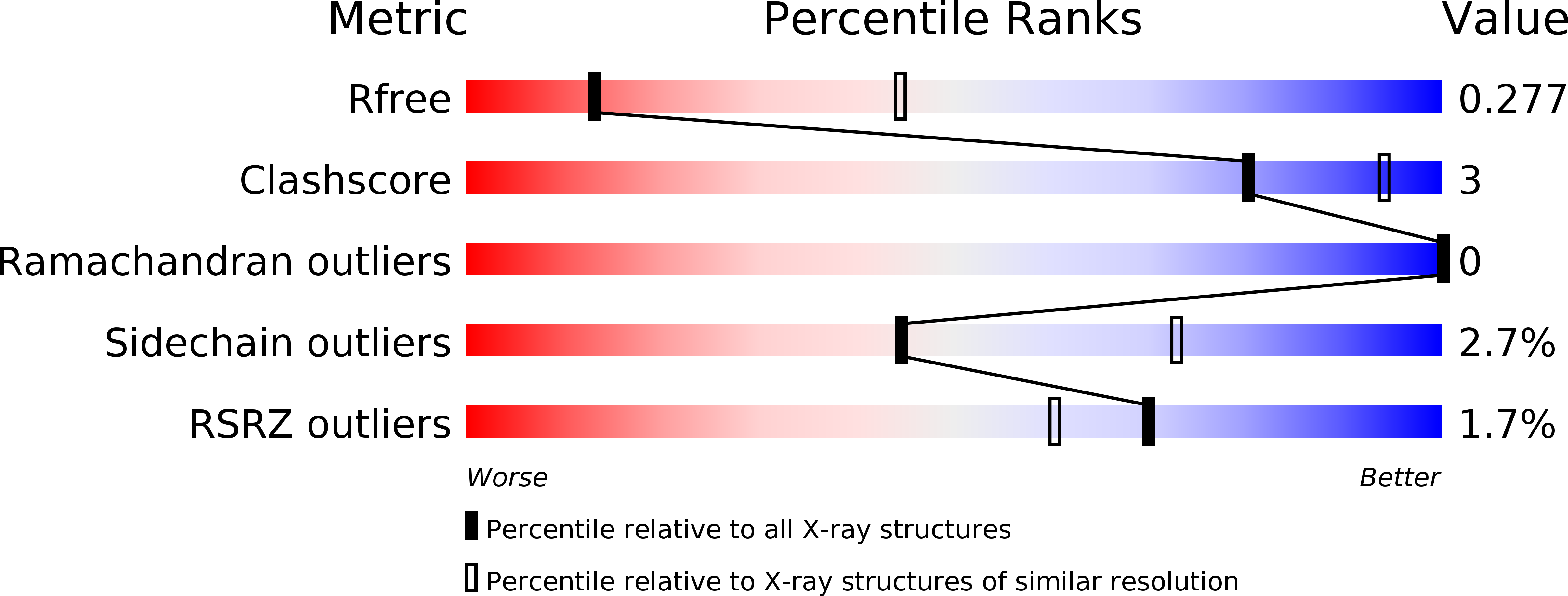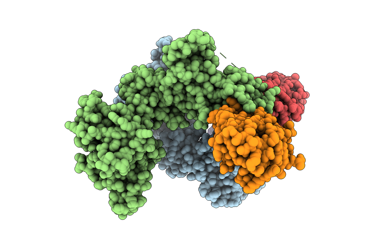
5UDH image
Entry Detail
PDB ID:
5UDH
Keywords:
Title:
HHARI/ARIH1-UBCH7~Ubiquitin
Biological Source:
PDB Version:
Deposition Date:
2016-12-27
Release Date:
2017-06-14
Method Details:
Experimental Method:
Resolution:
3.24 Å
R-Value Free:
0.27
R-Value Work:
0.22
R-Value Observed:
0.22
Space Group:
C 1 2 1
Macromolecular Entities
Polymer Type:polypeptide(L)
Description:E3 ubiquitin-protein ligase ARIH1
Chain IDs:A, C (auth: B)
Chain Length:470
Number of Molecules:2
Biological Source:Homo sapiens
Polymer Type:polypeptide(L)
Description:Ubiquitin-conjugating enzyme E2 L3
Mutations:C86K
Chain IDs:B (auth: C), D
Chain Length:156
Number of Molecules:2
Biological Source:Homo sapiens
Polymer Type:polypeptide(L)
Description:Ubiquitin C variant
Chain IDs:E
Chain Length:82
Number of Molecules:1
Biological Source:Homo sapiens
Ligand Molecules
Primary Citation
Structural Studies of HHARI/UbcH7Ub Reveal Unique E2Ub Conformational Restriction by RBR RING1.
Structure 25 890 900.e5 (2017)
PMID: 28552575 DOI: 10.1016/j.str.2017.04.013

Abstact

RING-between-RING (RBR) E3s contain RING1 domains that are structurally similar yet mechanistically distinct from canonical RING domains. Both types of E3 bind E2∼ubiquitin (E2∼Ub) via their RINGs but canonical RING E3s promote closed E2∼Ub conformations required for direct Ub transfer from the E2 to substrate, while RBR RING1s promote open E2∼Ub to favor Ub transfer to the E3 active site. This different RING/E2∼Ub conformation determines its direct target, which for canonical RING E3s is typically a substrate or substrate-linked Ub, but is the E3 active-site cysteine in the case of RBR-type E3s. Here we show that a short extension of HHARI RING1, namely Zn2+-loop II, not present in any RING E3s, acts as a steric wedge to disrupt closed E2∼Ub, providing a structural explanation for the distinctive RING1-dependent conformational restriction mechanism utilized by RBR E3s.

Legend

Protein

Chemical

Disease

Primary Citation of related structures