5UAN image
Deposition Date 2016-12-19
Release Date 2017-10-18
Last Version Date 2023-10-04
Entry Detail
PDB ID:
5UAN
Title:
Crystal structure of multi-domain RAR-beta-RXR-alpha heterodimer on DNA
Biological Source:
Source Organism:
Homo sapiens (Taxon ID: 9606)
Host Organism:
Method Details:
Experimental Method:
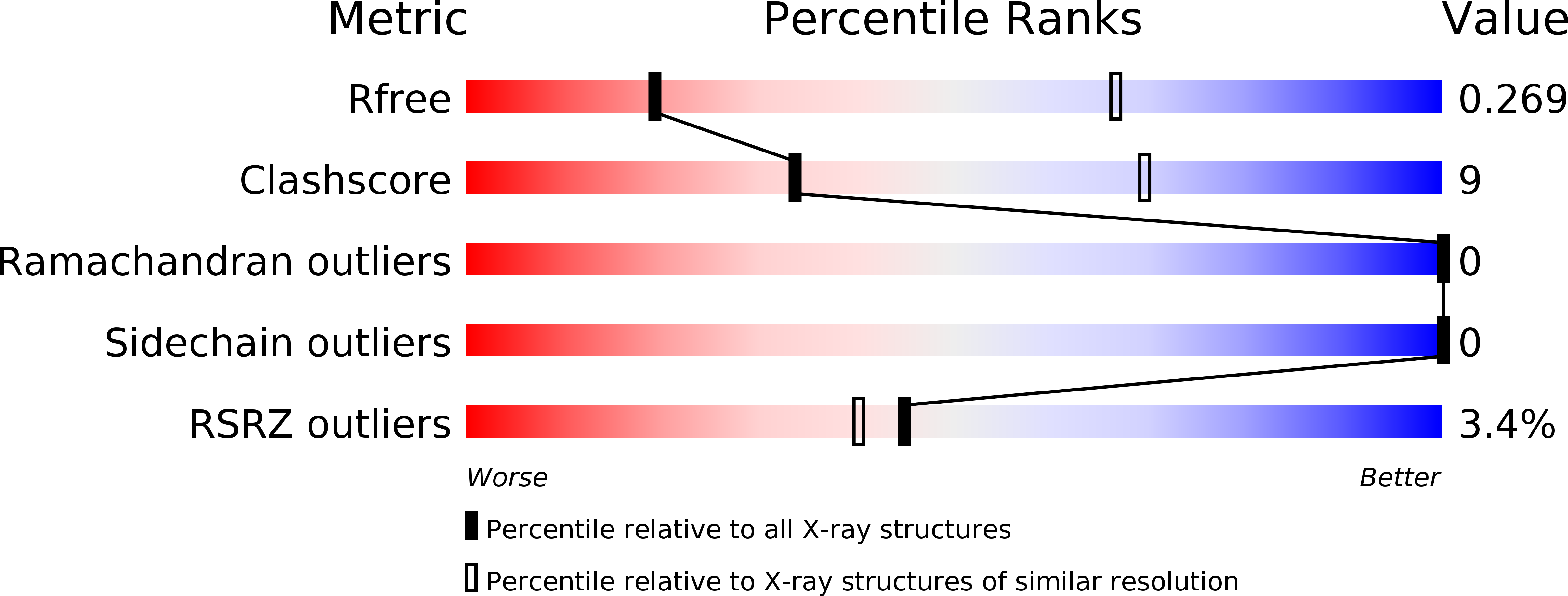
Resolution:
3.51 Å
R-Value Free:
0.27
R-Value Work:
0.22
R-Value Observed:
0.22
Space Group:
P 1 21 1
Macromolecular Entities
Structures with similar UniProt ID
Protein Blast
Polymer Type:polypeptide(L)
Molecule:Retinoic acid receptor RXR-alpha
Gene (Uniprot):RXRA
Chain IDs:A
Chain Length:365
Number of Molecules:1
Biological Source:Homo sapiens
Structures with similar UniProt ID
Protein Blast
Polymer Type:polypeptide(L)
Molecule:Retinoic acid receptor beta
Gene (Uniprot):RARB
Chain IDs:B
Chain Length:397
Number of Molecules:1
Biological Source:Homo sapiens
Structures with similar UniProt ID
Protein Blast
Polymer Type:polypeptide(L)
Molecule:Nuclear receptor coactivator 2
Chain IDs:C, D
Chain Length:10
Number of Molecules:2
Biological Source:Homo sapiens
Polymer Type:polydeoxyribonucleotide
Molecule:DNA (5'-D(*CP*TP*AP*GP*GP*TP*CP*AP*AP*AP*GP*GP*TP*CP*AP*GP*C)-3')
Chain IDs:E
Chain Length:17
Number of Molecules:1
Biological Source:Homo sapiens
Polymer Type:polydeoxyribonucleotide
Molecule:DNA (5'-D(*GP*CP*TP*GP*AP*CP*CP*TP*TP*TP*GP*AP*CP*CP*TP*AP*G)-3')
Chain IDs:F
Chain Length:17
Number of Molecules:1
Biological Source:Homo sapiens
Primary Citation
The quaternary architecture of RAR beta-RXR alpha heterodimer facilitates domain-domain signal transmission.
Nat Commun 8 868 868 (2017)
PMID: 29021580 DOI: 10.1038/s41467-017-00981-y

Abstact

Assessing the physical connections and allosteric communications in multi-domain nuclear receptor (NR) polypeptides has remained challenging, with few crystal structures available to show their overall structural organizations. Here we report the quaternary architecture of multi-domain retinoic acid receptor β-retinoic X receptor α (RARβ-RXRα) heterodimer bound to DNA, ligands and coactivator peptides, examined through crystallographic, hydrogen-deuterium exchange mass spectrometry, mutagenesis and functional studies. The RARβ ligand-binding domain (LBD) and DNA-binding domain (DBD) are physically connected to foster allosteric signal transmission between them. Direct comparisons among all the multi-domain NRs studied crystallographically to date show significant variations within their quaternary architectures, rather than a common architecture adhering to strict rules. RXR remains flexible and adaptive by maintaining loosely organized domains, while its heterodimerization partners use a surface patch on their LBDs to form domain-domain interactions with DBDs.Nuclear receptors (NR) are multidomain proteins, which makes their crystallization challenging. Here the authors present the crystal structure of the retinoic acid receptor β-retinoic X receptor α (RARβ-RXRα) heterodimer bound to DNA, ligands and coactivator peptides, which shows that NR quaternary architectures are variable.

Legend

Protein

Chemical

Disease

Primary Citation of related structures
Feedback Form
Name
Email
Institute
Feedback