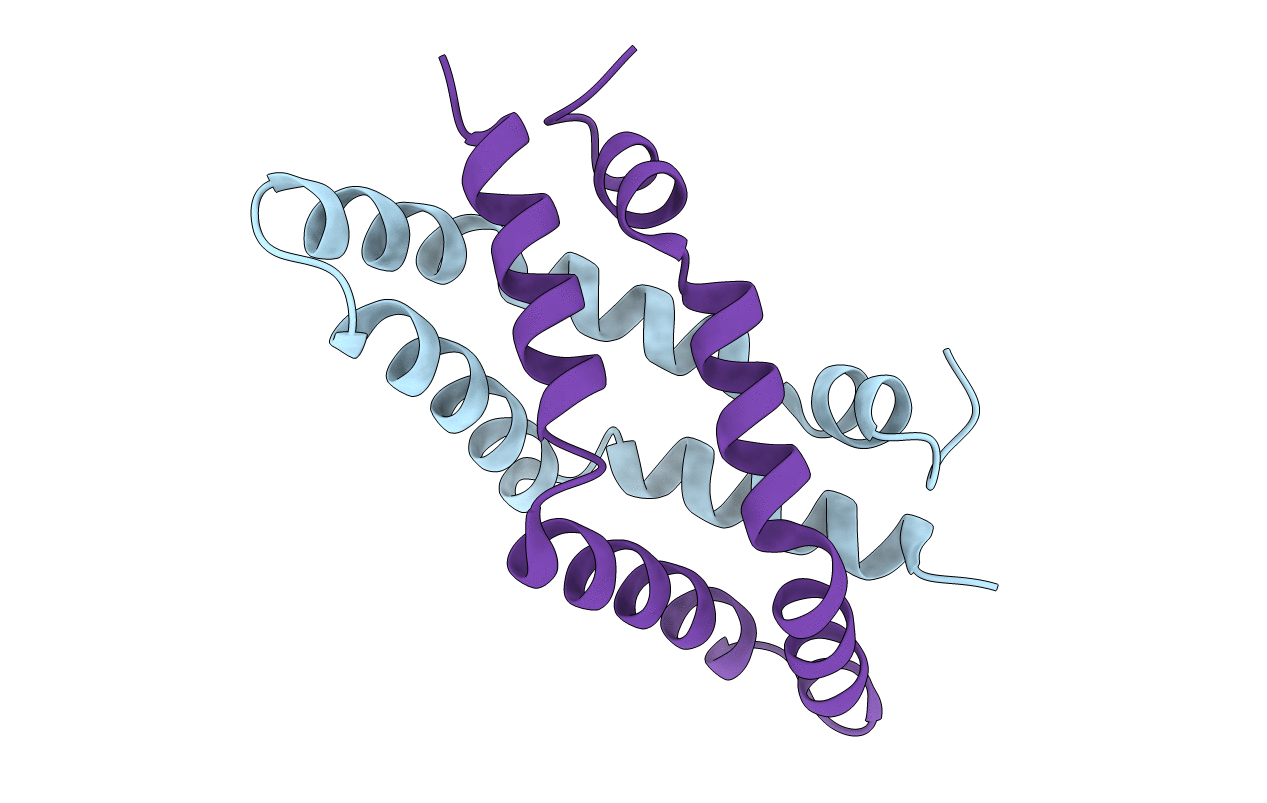
5U85 image
Deposition Date 2016-12-13
Release Date 2018-06-13
Last Version Date 2024-10-23
Entry Detail
PDB ID:
5U85
Title:
Murine saposin-D (SapD), open conformation
Biological Source:
Source Organism:
Mus musculus (Taxon ID: 10090)
Host Organism:
Method Details:
Experimental Method:
Resolution:
1.65 Å
R-Value Free:
0.18
R-Value Work:
0.16
R-Value Observed:
0.16
Space Group:
P 21 21 21
Macromolecular Entities
Structures with similar UniProt ID
Protein Blast
Polymer Type:polypeptide(L)
Molecule:Saposin-D
Gene (Uniprot):Psap
Chain IDs:A, B
Chain Length:83
Number of Molecules:2
Biological Source:Mus musculus
Primary Citation
Crystal structure of saposin D in an open conformation.
J. Struct. Biol. 204 145 150 (2018)
PMID: 30026085 DOI: 10.1016/j.jsb.2018.07.011

Abstact

Saposins are accessory proteins that aid in the degradation of sphingolipids by hydrolytic enzymes. Their structure usually comprises four α-helices arranged in various conformations including an open, V-shaped form that is generally associated with the ability to interact with membranes and/or enzymes to accentuate activity. Saposin D is required by the lysosomal hydrolase, acid ceramidase, which breaks down ceramide into sphingosine and free fatty acid, to display optimal activity. The structure of saposin D was previously determined in an inactive conformation, revealing a monomeric, closed and compact form. Here, we present the crystal structure of the open, V-shaped form of saposin D. The overall shape is similar to the open conformation found in other saposins with slight differences in the angles between the α-helices. The structure forms a dimer that serves to stabilize the hydrophobic surface exposed in the open form, which results in an internal, hydrophobic cavity that could be used to carry extracted membrane lipids.

Legend

Protein

Chemical

Disease

Primary Citation of related structures