5U52 image
Deposition Date 2016-12-06
Release Date 2017-05-24
Last Version Date 2024-10-16
Entry Detail
PDB ID:
5U52
Keywords:
Title:
2 helix minimized version of the B-domain from Protein A (Z34C0 bound to IgG1 Fc (monoclinic form)
Biological Source:
Source Organism:
Host Organism:
Method Details:
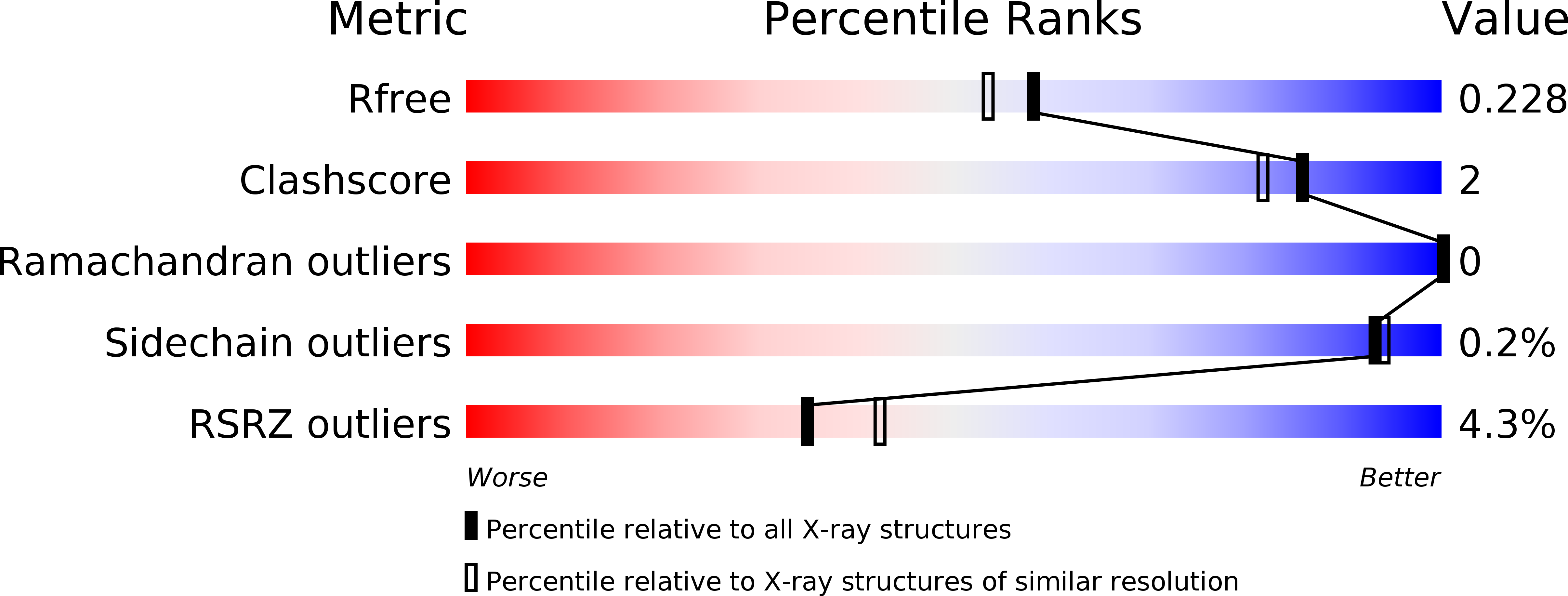
Experimental Method:
Resolution:
1.94 Å
R-Value Free:
0.22
R-Value Work:
0.19
R-Value Observed:
0.19
Space Group:
P 1 21 1
Macromolecular Entities
Structures with similar UniProt ID
Protein Blast
Polymer Type:polypeptide(L)
Molecule:IGG1 FC
Chain IDs:A, B
Chain Length:209
Number of Molecules:2
Biological Source:Homo sapiens
Polymer Type:polypeptide(L)
Molecule:Mini Z domain
Chain IDs:C (auth: E), D (auth: Z)
Chain Length:34
Number of Molecules:2
Biological Source:Staphylococcus aureus
Primary Citation
3-2-1: Structural insights from stepwise shrinkage of a three-helix Fc-binding domain to a single helix.
Protein Eng. Des. Sel. 30 619 625 (2017)
PMID: 28475752 DOI: 10.1093/protein/gzx029

Abstact

The well-studied B-domain from Staphylococcal protein A is a 59 amino acid three-helix bundle that binds the Fc portion of IgG with a dissociation constant of ~35 nM. The B-domain variant bearing a Gly to Ala mutation (=Z-domain) has been the subject of efforts to minimize a domain's size while retaining its function. We report X-ray crystallographic characterization of three steps in such a process using complexes with Fc: the full three-helix Z-domain, a 34 amino acid two-helix version called Z34C and a 13 amino acid single helix stabilized with an exo-helix tether, called LH1.

Legend

Protein

Chemical

Disease

Primary Citation of related structures