Deposition Date
2016-12-02
Release Date
2017-02-01
Last Version Date
2024-03-06
Entry Detail
PDB ID:
5U3G
Keywords:
Title:
Structure of the Dickeya dadantii ykkC riboswitch bound to guanidinium
Biological Source:
Source Organism(s):
Dickeya dadantii (Taxon ID: 204038)
Method Details:
Experimental Method:
Resolution:
2.30 Å
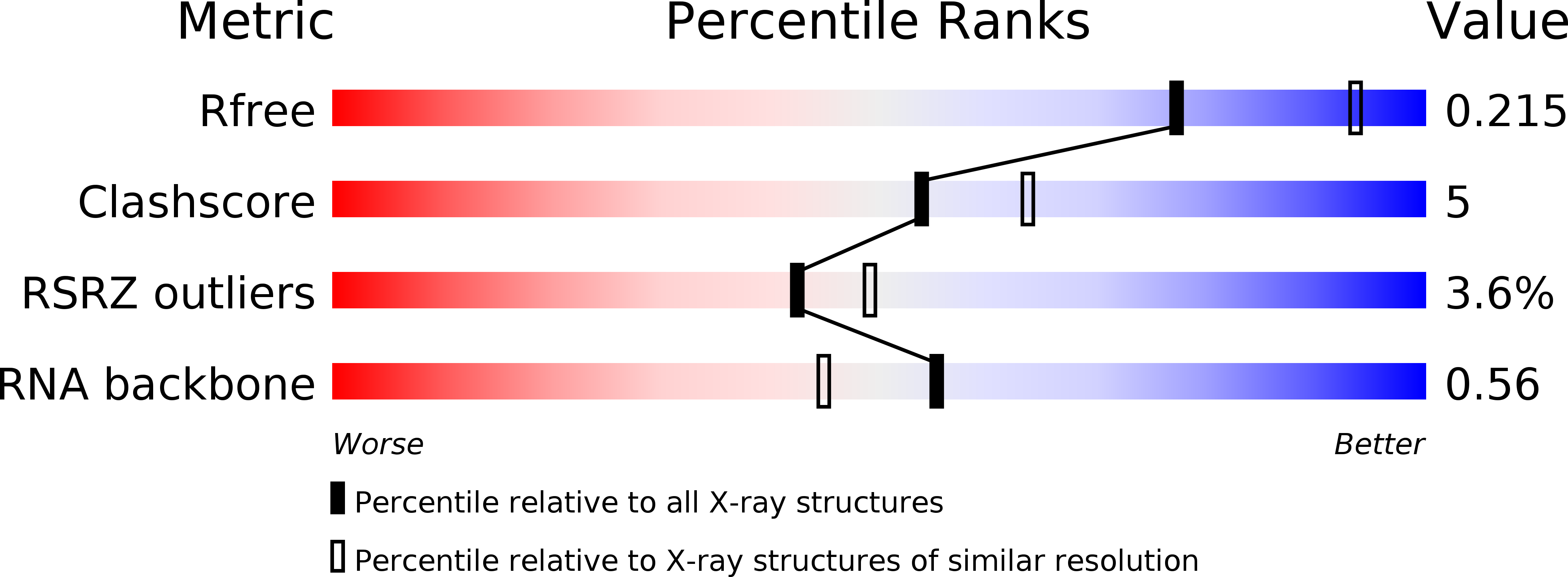
R-Value Free:
0.21
R-Value Work:
0.19
R-Value Observed:
0.19
Space Group:
I 2 2 2