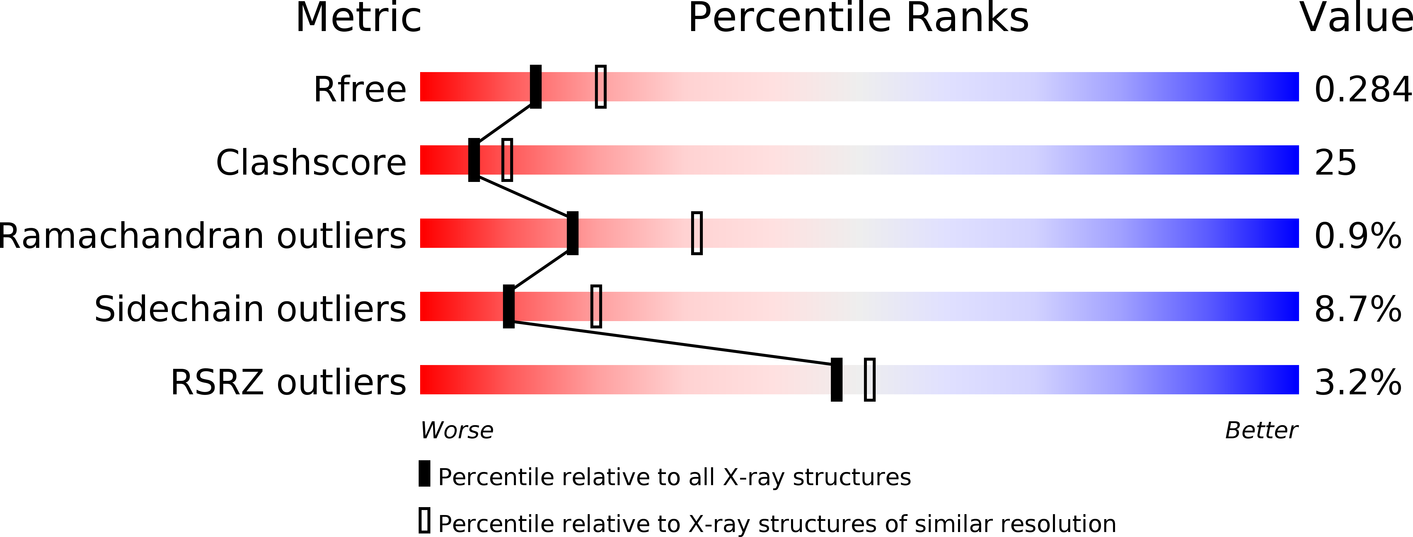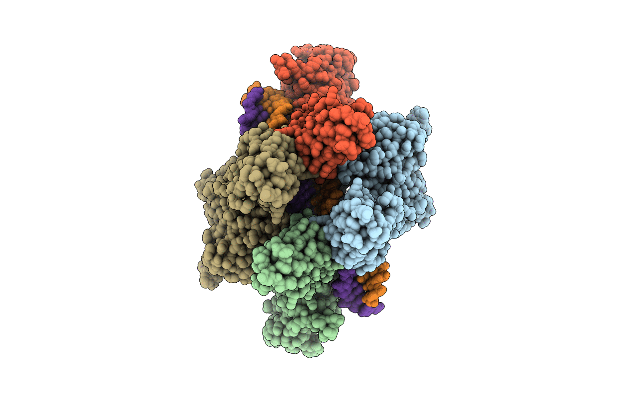
5U01 image
Deposition Date 2016-11-22
Release Date 2017-09-27
Last Version Date 2023-10-04
Entry Detail
PDB ID:
5U01
Title:
Cooperative DNA binding by two RelA dimers
Biological Source:
Source Organism:
Synthesium (Taxon ID: 638370)
Mus musculus (Taxon ID: 10090)
Method Details:
Experimental Method:
Resolution:
2.50 Å
R-Value Free:
0.27
R-Value Work:
0.22
R-Value Observed:
0.22
Space Group:
P 1 21 1
Macromolecular Entities
Polymer Type:polypeptide(L)
Molecule:Transcription factor p65
Gene (Uniprot):Rela
Chain IDs:A, B, C, D
Chain Length:273
Number of Molecules:4
Biological Source:Mus musculus
Polymer Type:polydeoxyribonucleotide
Molecule:DNA (27-MER)
Chain IDs:E (auth: F), F (auth: E)
Chain Length:27
Number of Molecules:2
Biological Source:Synthesium
Primary Citation
DNA-binding affinity and transcriptional activity of the RelA homodimer of nuclear factor kappa B are not correlated.
J. Biol. Chem. 292 18821 18830 (2017)
PMID: 28935669 DOI: 10.1074/jbc.M117.813980

Abstact

The nuclear factor κB (NF-κB) transcription factor family regulates genes involved in cell proliferation and inflammation. The promoters of these genes often contain NF-κB-binding sites (κB sites) arranged in tandem. How NF-κB activates transcription through these multiple sites is incompletely understood. We report here an X-ray crystal structure of homodimers comprising the RelA DNA-binding domain containing the Rel homology region (RHR) in NF-κB bound to an E-selectin promoter fragment with tandem κB sites. This structure revealed that two dimers bind asymmetrically to the symmetrically arranged κB sites at which multiple cognate contacts between one dimer to the corresponding DNA are broken. Because simultaneous RelA-RHR dimer binding to tandem sites in solution was anti-cooperative, we inferred that asymmetric RelA-RHR binding with fewer contacts likely indicates a dissociative binding mode. We found that both κB sites are essential for reporter gene activation by full-length RelA homodimer, suggesting that dimers facilitate DNA binding to each other even though their stable co-occupation is not promoted. Promoter variants with altered spacing and orientation of tandem κB sites displayed unexpected reporter activities that were not explained by the solution-binding pattern of RelA-RHR. Remarkably, full-length RelA bound all DNAs with a weaker affinity and specificity. Moreover, the transactivation domain played a negative role in DNA binding. These observations suggest that other nuclear factors influence full-length RelA binding to DNA by neutralizing the transactivation domain negative effect. We propose that DNA binding by NF-κB dimers is highly complex and modulated by facilitated association-dissociation processes.

Legend

Protein

Chemical

Disease

Primary Citation of related structures