Deposition Date
2016-11-16
Release Date
2017-10-25
Last Version Date
2024-10-23
Entry Detail
PDB ID:
5TXF
Keywords:
Title:
Crystal structure of Lecithin:cholesterol acyltransferase (LCAT) in a closed conformation
Biological Source:
Source Organism(s):
Homo sapiens (Taxon ID: 9606)
Expression System(s):
Method Details:
Experimental Method:
Resolution:
3.10 Å
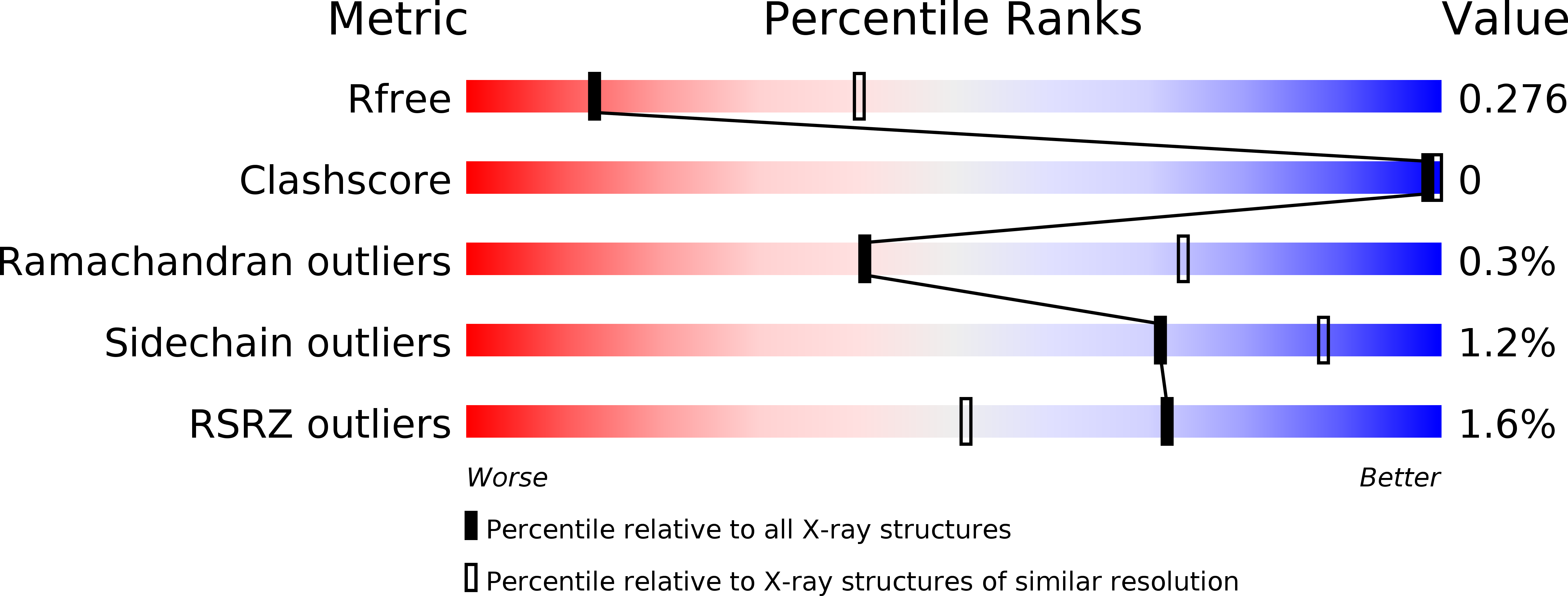
R-Value Free:
0.26
R-Value Work:
0.24
R-Value Observed:
0.25
Space Group:
P 1 21 1