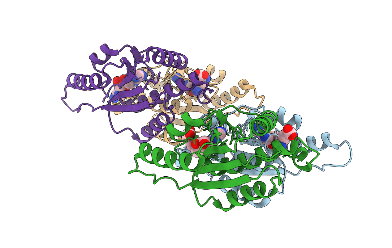
5TWK image
Entry Detail
PDB ID:
5TWK
Title:
Crystal Structure of RlmH in Complex with Sinefungin
Biological Source:
Source Organism:
Host Organism:
PDB Version:
Deposition Date:
2016-11-14
Release Date:
2017-05-03
Method Details:
Experimental Method:
Resolution:
2.10 Å
R-Value Free:
0.23
R-Value Work:
0.18
R-Value Observed:
0.18
Space Group:
P 1 21 1
Macromolecular Entities
Polymer Type:polypeptide(L)
Description:Ribosomal RNA large subunit methyltransferase H
Chain IDs:A (auth: C), B (auth: D), C (auth: A), D (auth: B)
Chain Length:162
Number of Molecules:4
Biological Source:Escherichia coli
Ligand Molecules
Primary Citation
Small methyltransferase RlmH assembles a composite active site to methylate a ribosomal pseudouridine.
Sci Rep 7 969 969 (2017)
PMID: 28428565 DOI: 10.1038/s41598-017-01186-5

Abstact

Eubacterial ribosomal large-subunit methyltransferase H (RlmH) methylates 23S ribosomal RNA pseudouridine 1915 (Ψ1915), which lies near the ribosomal decoding center. The smallest member of the SPOUT superfamily of methyltransferases, RlmH lacks the RNA recognition domain found in larger methyltransferases. The catalytic mechanism of RlmH enzyme is unknown. Here, we describe the structures of RlmH bound to S-adenosyl-methionine (SAM) and the methyltransferase inhibitor sinefungin. Our structural and biochemical studies reveal catalytically essential residues in the dimer-mediated asymmetrical active site. One monomer provides the SAM-binding site, whereas the conserved C-terminal tail of the second monomer provides residues essential for catalysis. Our findings elucidate the mechanism by which a small protein dimer assembles a functionally asymmetric architecture.

Legend

Protein

Chemical

Disease

Primary Citation of related structures