5TVM image
Deposition Date 2016-11-09
Release Date 2017-01-11
Last Version Date 2024-11-13
Entry Detail
PDB ID:
5TVM
Keywords:
Title:
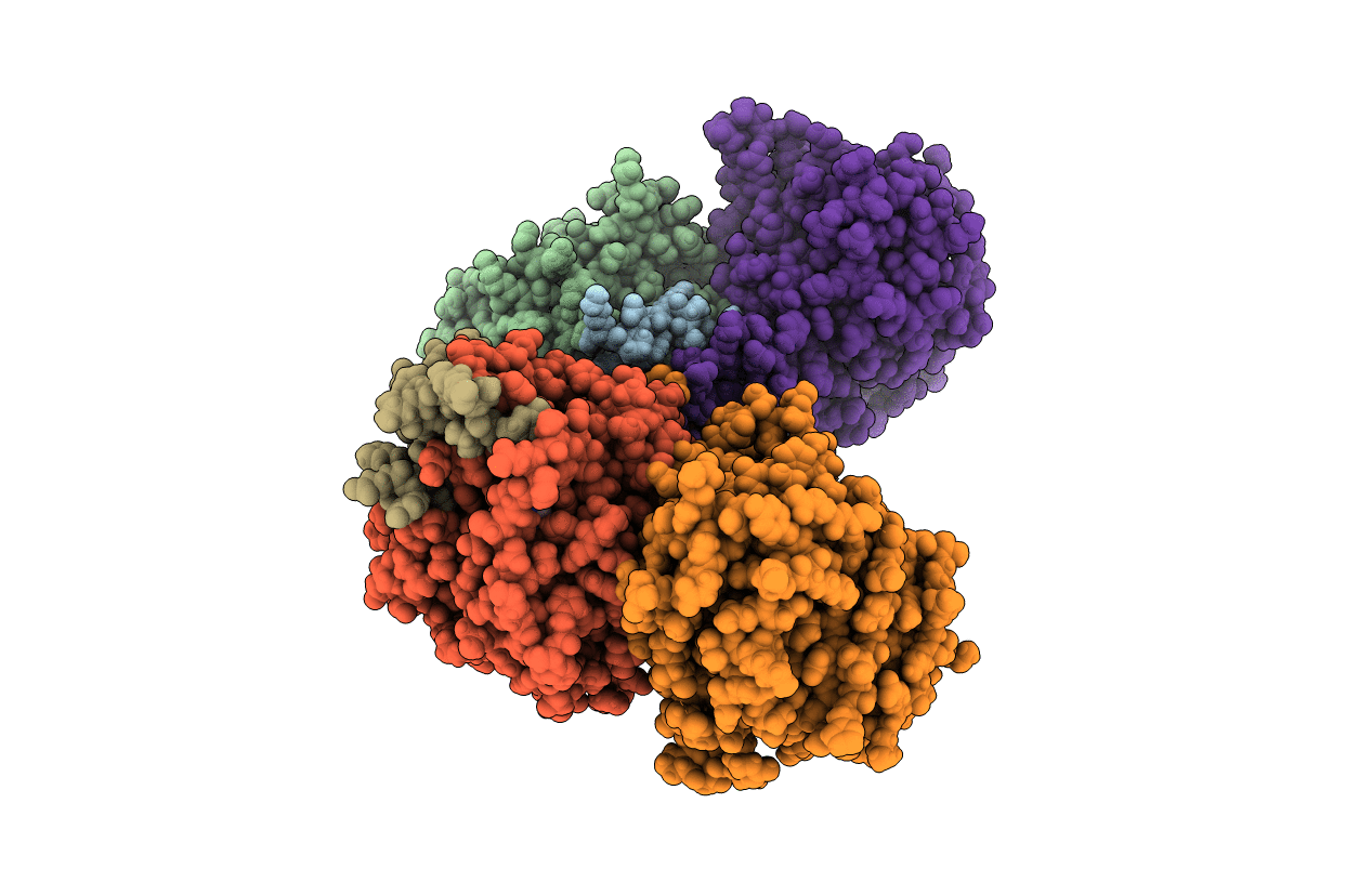
Crystal structure of Trypanosoma brucei AdoMetDC/prozyme heterodimer
Biological Source:
Host Organism:
Method Details:
Experimental Method:
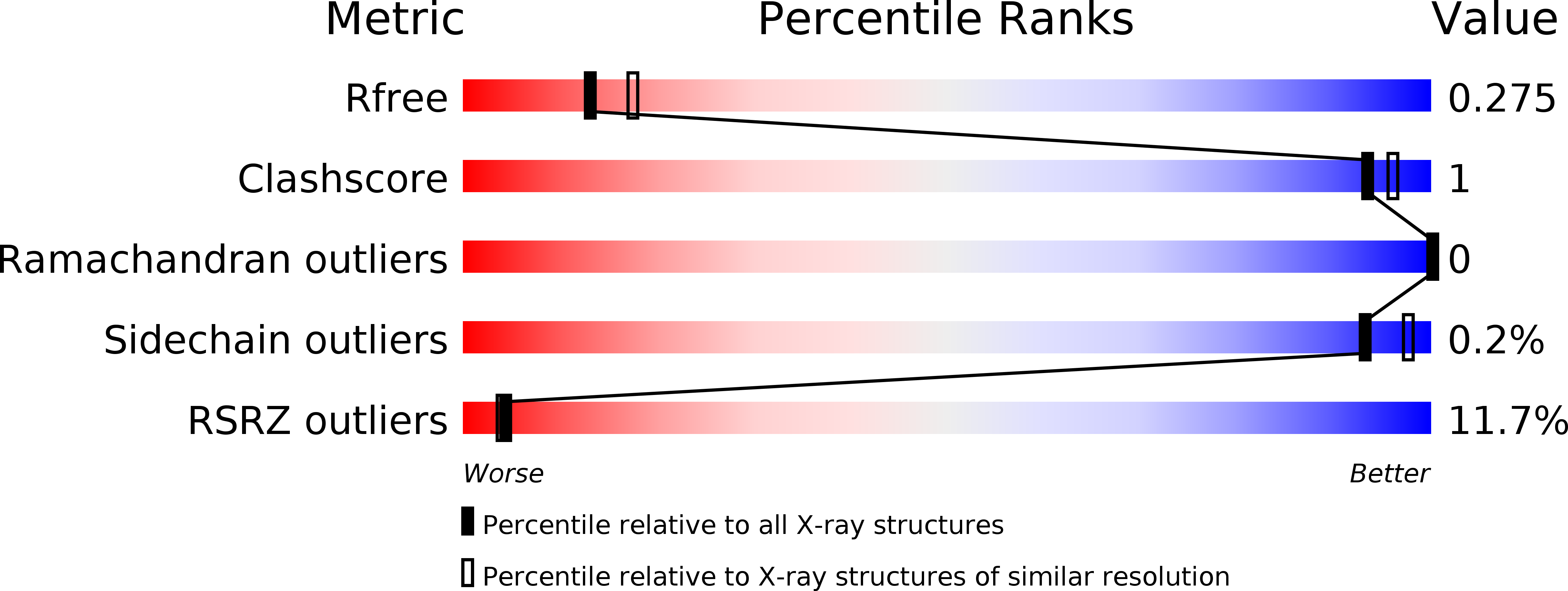
Resolution:
2.41 Å
R-Value Free:
0.27
R-Value Work:
0.22
R-Value Observed:
0.23
Space Group:
P 1 21 1
Macromolecular Entities
Structures with similar UniProt ID
Protein Blast
Polymer Type:polypeptide(L)
Molecule:S-adenosylmethionine decarboxylase beta chain
Gene (Uniprot):Tb06.26G9.820
Chain IDs:A, C
Chain Length:85
Number of Molecules:2
Biological Source:Trypanosoma brucei brucei (strain 927/4 GUTat10.1)
Structures with similar UniProt ID
Protein Blast
Polymer Type:polypeptide(L)
Molecule:S-adenosylmethionine decarboxylase alpha chain
Gene (Uniprot):Tb06.26G9.820
Chain IDs:B, D
Chain Length:285
Number of Molecules:2
Biological Source:Trypanosoma brucei brucei (strain 927/4 GUTat10.1)
Structures with similar UniProt ID
Protein Blast
Polymer Type:polypeptide(L)
Molecule:S-adenosylmethionine decarboxylase proenzyme-like, putative
Gene (Uniprot):Tb06.26G9.750
Chain IDs:E, F
Chain Length:325
Number of Molecules:2
Biological Source:Trypanosoma brucei brucei (strain 927/4 GUTat10.1)
Primary Citation
Relief of autoinhibition by conformational switch explains enzyme activation by a catalytically dead paralog.
Elife 5 ? ? (2016)
PMID: 27977001 DOI: 10.7554/eLife.20198

Abstact

Catalytically inactive enzyme paralogs occur in many genomes. Some regulate their active counterparts but the structural principles of this regulation remain largely unknown. We report X-ray structures of Trypanosoma brucei S-adenosylmethionine decarboxylase alone and in functional complex with its catalytically dead paralogous partner, prozyme. We show monomeric TbAdoMetDC is inactive because of autoinhibition by its N-terminal sequence. Heterodimerization with prozyme displaces this sequence from the active site through a complex mechanism involving a cis-to-trans proline isomerization, reorganization of a β-sheet, and insertion of the N-terminal α-helix into the heterodimer interface, leading to enzyme activation. We propose that the evolution of this intricate regulatory mechanism was facilitated by the acquisition of the dimerization domain, a single step that can in principle account for the divergence of regulatory schemes in the AdoMetDC enzyme family. These studies elucidate an allosteric mechanism in an enzyme and a plausible scheme by which such complex cooperativity evolved.

Legend

Protein

Chemical

Disease

Primary Citation of related structures