
Deposition Date
2016-10-25
Release Date
2017-02-08
Last Version Date
2024-11-13
Entry Detail
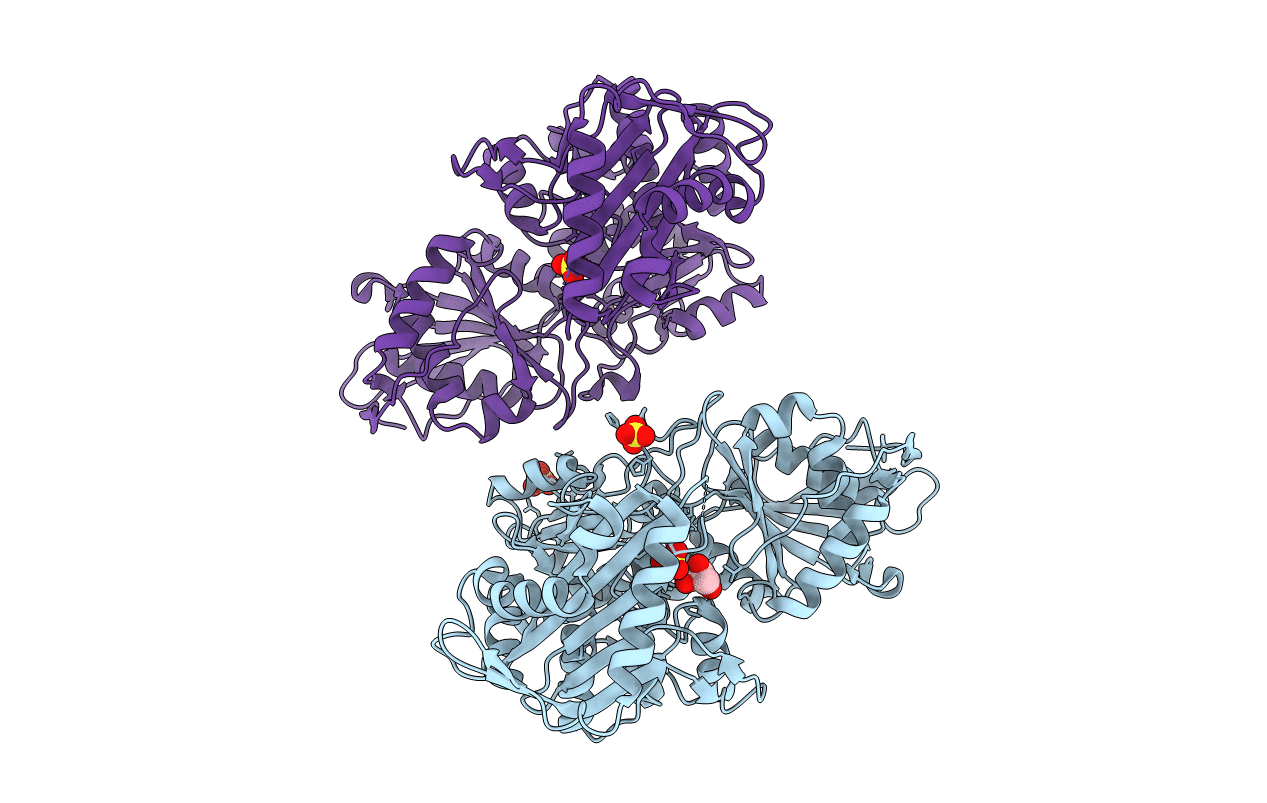
PDB ID:
5TR2
Keywords:
Title:
Crystal structure of the D263G missense variant of human PGM1
Biological Source:
Source Organism(s):
Homo sapiens (Taxon ID: 9606)
Expression System(s):
Method Details:
Experimental Method:
Resolution:
2.50 Å
R-Value Free:
0.29
R-Value Work:
0.22
R-Value Observed:
0.22
Space Group:
P 41 21 2
