5TLC image
Entry Detail
PDB ID:
5TLC
Keywords:
Title:
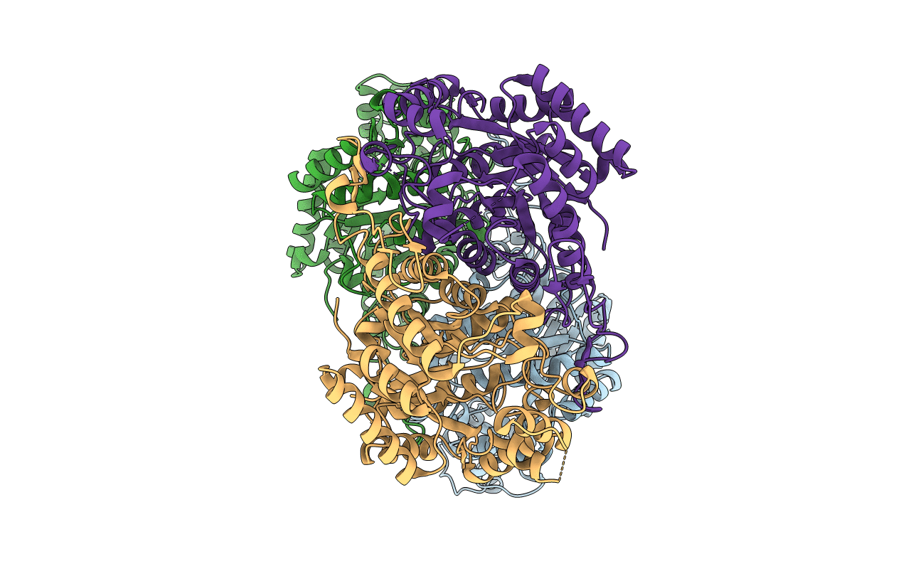
Crystal structure of BdsA from Bacillus subtilis WU-S2B
Biological Source:
Source Organism:
Host Organism:
PDB Version:
Deposition Date:
2016-10-11
Release Date:
2017-05-03
Method Details:
Experimental Method:
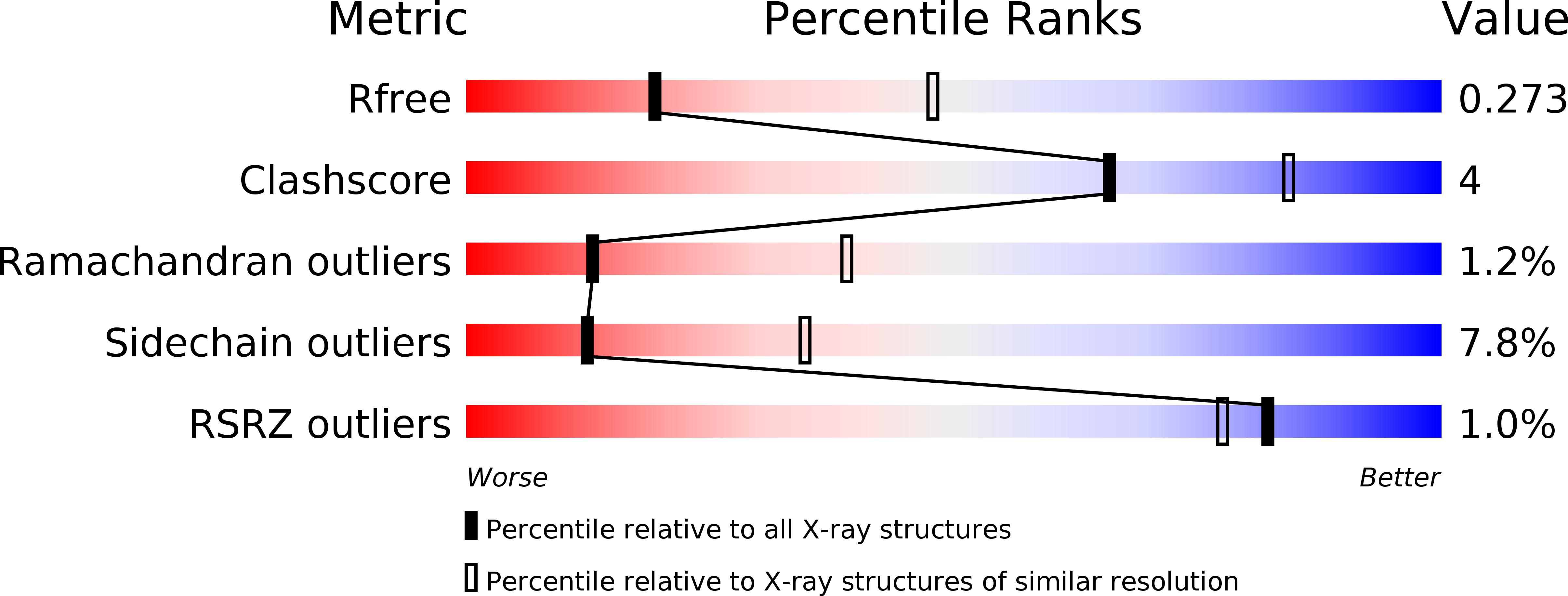
Resolution:
2.80 Å
R-Value Free:
0.27
R-Value Work:
0.21
R-Value Observed:
0.21
Space Group:
P 32
Macromolecular Entities
Polymer Type:polypeptide(L)
Description:Dibenzothiophene desulfurization enzyme A
Chain IDs:A, B, C, D
Chain Length:453
Number of Molecules:4
Biological Source:Bacillus subtilis
Primary Citation
Crystal structure of dibenzothiophene sulfone monooxygenase BdsA from Bacillus subtilis WU-S2B
Proteins 85 1171 1177 (2017)
PMID: 28205250 DOI: 10.1002/prot.25267

Abstact

The dibenzothiophene (DBT) sulfone monooxygenase BdsA from Bacillus subtilis WU-S2B catalyzes the conversion of DBT sulfone to 2'-hydroxybiphenyl 2-sulfinate. We report the crystal structures of BdsA at a resolution of 2.80 Å. BdsA exists as a homotetramer with a dimer-of-dimers configuration in the crystal, and the interaction between E288 and R296 in BdsA is important for tetramer formation. A structural comparison with homologous proteins shows that the orientation and location of the α9-α12 helices in BdsA are closer to those of the closed form than those of the open form in the EDTA monooxygenase EmoA. Proteins 2017; 85:1171-1177. © 2017 Wiley Periodicals, Inc.

Legend

Protein

Chemical

Disease

Primary Citation of related structures