5TK5 image
Deposition Date 2016-10-06
Release Date 2017-04-12
Last Version Date 2023-10-04
Entry Detail
PDB ID:
5TK5
Keywords:
Title:
Crystal structure of human 3HAO with iron bound in the active site
Biological Source:
Source Organism:
Homo sapiens (Taxon ID: 9606)
Host Organism:
Method Details:
Experimental Method:
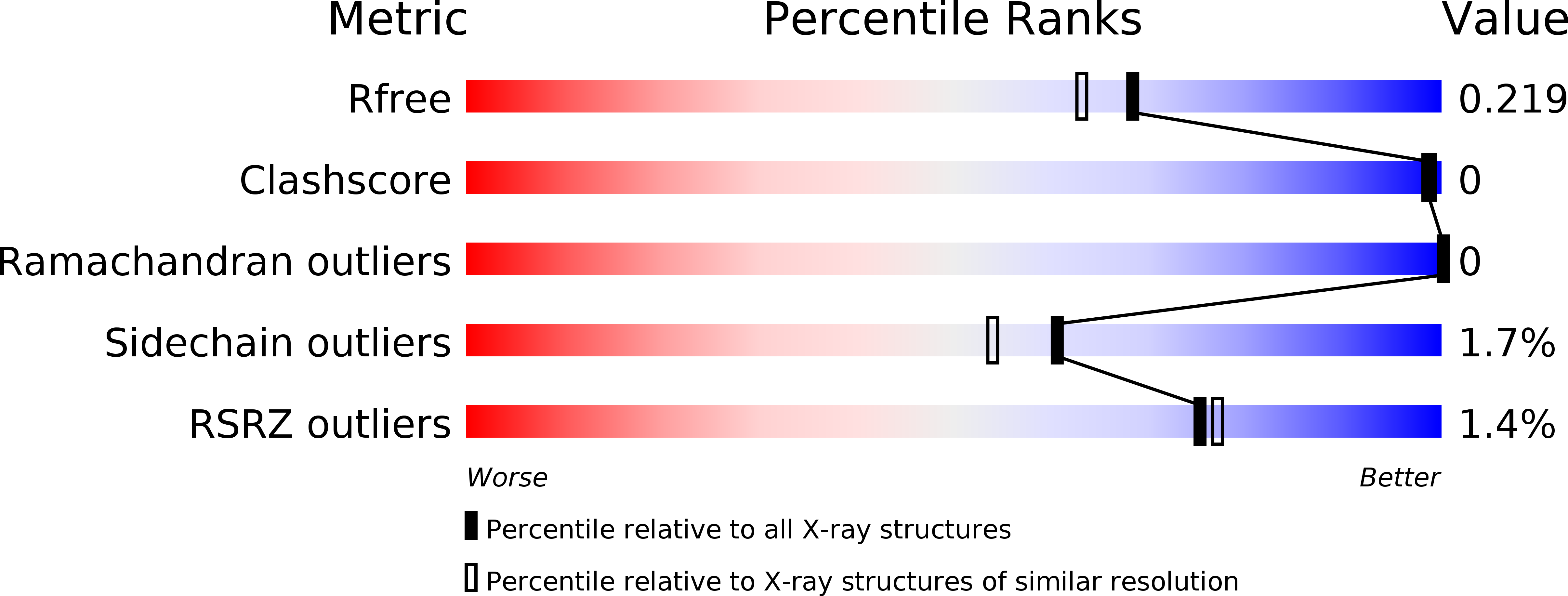
Resolution:
1.88 Å
R-Value Free:
0.21
R-Value Work:
0.20
R-Value Observed:
0.20
Space Group:
P 65 2 2
Macromolecular Entities
Polymer Type:polypeptide(L)
Molecule:3-hydroxyanthranilate 3,4-dioxygenase
Gene (Uniprot):HAAO
Chain IDs:A
Chain Length:290
Number of Molecules:1
Biological Source:Homo sapiens
Primary Citation
Crystal structures of human 3-hydroxyanthranilate 3,4-dioxygenase with native and non-native metals bound in the active site.
Acta Crystallogr D Struct Biol 73 340 348 (2017)
PMID: 28375145 DOI: 10.1107/S2059798317002029

Abstact

3-Hydroxyanthranilate 3,4-dioxygenase (3HAO) is an enzyme in the microglial branch of the kynurenine pathway of tryptophan degradation. 3HAO is a non-heme iron-containing, ring-cleaving extradiol dioxygenase that catalyzes the addition of both atoms of O2 to the kynurenine pathway metabolite 3-hydroxyanthranilic acid (3-HANA) to form quinolinic acid (QUIN). QUIN is a highly potent excitotoxin that has been implicated in a number of neurodegenerative conditions, making 3HAO a target for pharmacological downregulation. Here, the first crystal structure of human 3HAO with the native iron bound in its active site is presented, together with an additional structure with zinc (a known inhibitor of human 3HAO) bound in the active site. The metal-binding environment is examined both structurally and via inductively coupled plasma mass spectrometry (ICP-MS), X-ray fluorescence spectroscopy (XRF) and electron paramagnetic resonance spectroscopy (EPR). The studies identified Met35 as the source of potential new interactions with substrates and inhibitors, which may prove useful in future therapeutic efforts.

Legend

Protein

Chemical

Disease

Primary Citation of related structures