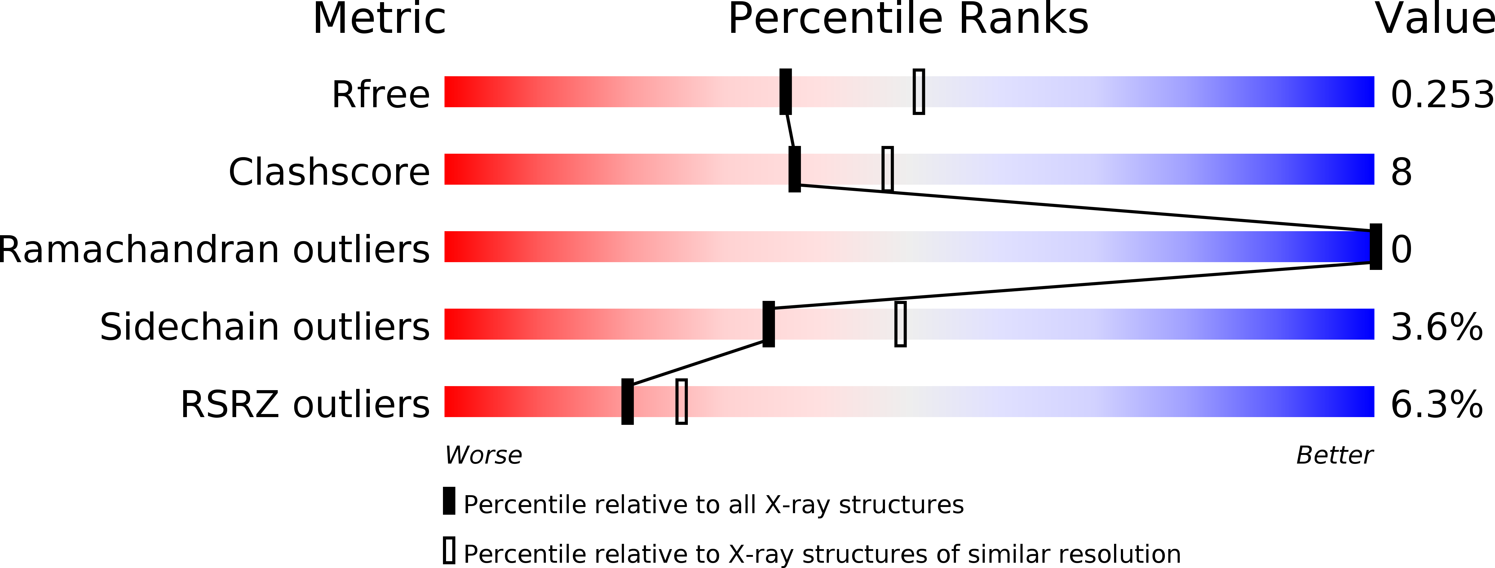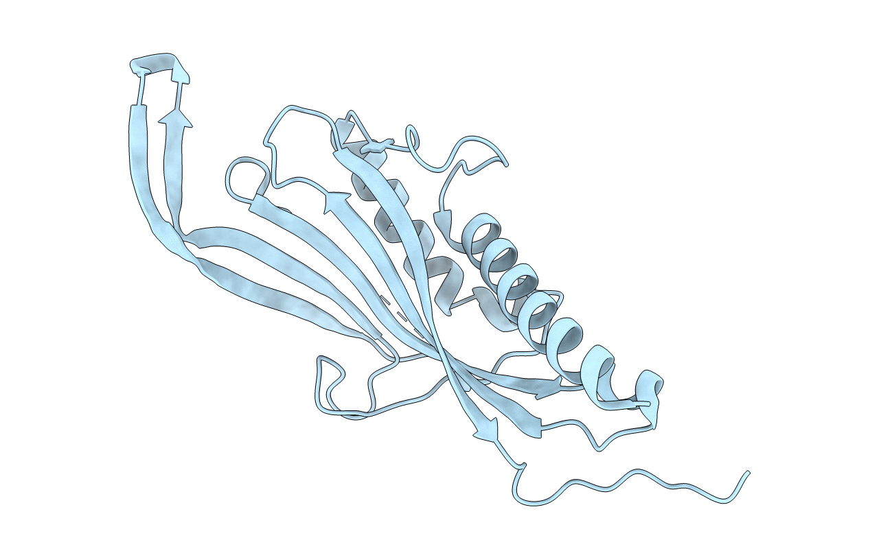
5TJA image
Deposition Date 2016-10-04
Release Date 2017-01-25
Last Version Date 2024-10-23
Entry Detail
PDB ID:
5TJA
Title:
I-II linker of TRPML1 channel at pH 6
Biological Source:
Source Organism:
Homo sapiens (Taxon ID: 9606)
Host Organism:
Method Details:
Experimental Method:
Resolution:
2.30 Å
R-Value Free:
0.25
R-Value Work:
0.22
Space Group:
I 4 2 2
Macromolecular Entities
Structures with similar UniProt ID
Protein Blast
Polymer Type:polypeptide(L)
Molecule:Mucolipin-1
Gene (Uniprot):MCOLN1
Chain IDs:A
Chain Length:223
Number of Molecules:1
Biological Source:Homo sapiens
Primary Citation
Structural basis of dual Ca(2+)/pH regulation of the endolysosomal TRPML1 channel.
Nat. Struct. Mol. Biol. 24 205 213 (2017)
PMID: 28112729 DOI: 10.1038/nsmb.3362

Abstact

The activities of organellar ion channels are often regulated by Ca2+ and H+, which are present in high concentrations in many organelles. Here we report a structural element critical for dual Ca2+/pH regulation of TRPML1, a Ca2+-release channel crucial for endolysosomal function. TRPML1 mutations cause mucolipidosis type IV (MLIV), a severe lysosomal storage disorder characterized by neurodegeneration, mental retardation and blindness. We obtained crystal structures of the 213-residue luminal domain of human TRPML1 containing three missense MLIV-causing mutations. This domain forms a tetramer with a highly electronegative central pore formed by a novel luminal pore loop. Cysteine cross-linking and cryo-EM analyses confirmed that this architecture occurs in the full-length channel. Structure-function studies demonstrated that Ca2+ and H+ interact with the luminal pore and exert physiologically important regulation. The MLIV-causing mutations disrupt the luminal-domain structure and cause TRPML1 mislocalization. Our study reveals the structural underpinnings of TRPML1's regulation, assembly and pathogenesis.

Legend

Protein

Chemical

Disease

Primary Citation of related structures