5TH3 image
Entry Detail
PDB ID:
5TH3
Title:
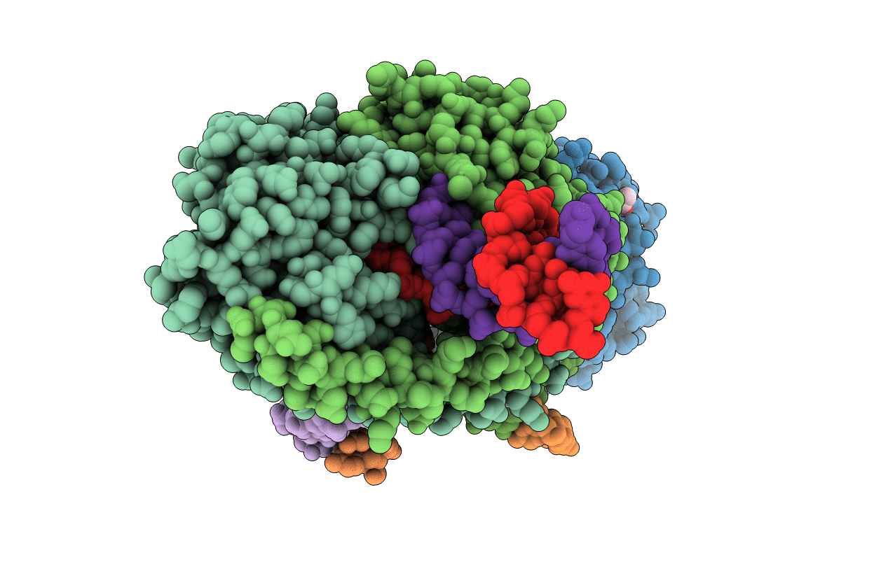
Restriction/modification system-Type II R.SwaI cleaved DNA complex
Biological Source:
PDB Version:
Deposition Date:
2016-09-29
Release Date:
2016-12-21
Method Details:
Experimental Method:
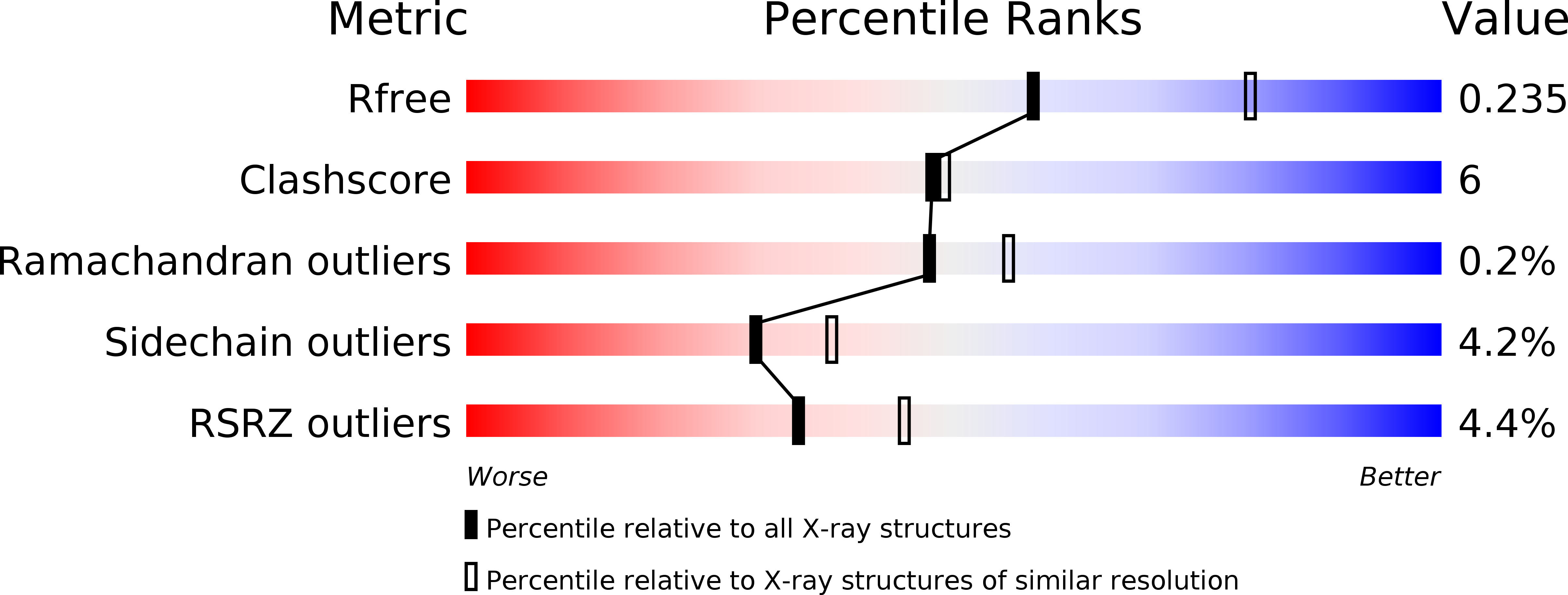
Resolution:
2.33 Å
R-Value Free:
0.23
R-Value Work:
0.19
R-Value Observed:
0.20
Space Group:
P 1 21 1
Macromolecular Entities
Polymer Type:polypeptide(L)
Description:R-SwaI protein
Chain IDs:A, B, C, D
Chain Length:226
Number of Molecules:4
Biological Source:Staphylococcus warneri
Polymer Type:polydeoxyribonucleotide
Description:DNA (cleaved 25-MER, portion 1)
Chain IDs:E (auth: H), I (auth: J)
Chain Length:13
Number of Molecules:2
Biological Source:synthetic construct
Polymer Type:polydeoxyribonucleotide
Description:DNA (cleaved 26-MER, portion 1)
Chain IDs:G (auth: I), K
Chain Length:14
Number of Molecules:2
Biological Source:synthetic construct
Polymer Type:polydeoxyribonucleotide
Description:DNA (cleaved 25-MER, portion 2)
Chain IDs:F (auth: h), J (auth: j)
Chain Length:13
Number of Molecules:2
Biological Source:synthetic construct
Polymer Type:polydeoxyribonucleotide
Description:DNA (cleaved 26-MER, portion 2)
Chain IDs:H (auth: i), L (auth: k)
Chain Length:14
Number of Molecules:2
Biological Source:synthetic construct
Primary Citation
DNA recognition by the SwaI restriction endonuclease involves unusual distortion of an 8 base pair A:T-rich target.
Nucleic Acids Res. 45 1516 1528 (2017)
PMID: 28180307 DOI: 10.1093/nar/gkw1200

Abstact

R.SwaI, a Type IIP restriction endonuclease, recognizes a palindromic eight base pair (bp) symmetric sequence, 5΄-ATTTAAAT-3΄, and cleaves that target at its center to generate blunt-ended DNA fragments. Here, we report three crystal structures of SwaI: unbound enzyme, a DNA-bound complex with calcium ions; and a DNA-bound, fully cleaved complex with magnesium ions. We compare these structures to two structurally similar ‘PD-D/ExK’ restriction endonucleases (EcoRV and HincII) that also generate blunt-ended products, and to a structurally distinct enzyme (the HNH endonuclease PacI) that also recognizes an 8-bp target site consisting solely of A:T base pairs. Binding by SwaI induces an extreme bend in the target sequence accompanied by un-pairing and re-ordering of its central A:T base pairs. This result is reminiscent of a more dramatic target deformation previously described for PacI, implying that long A:T-rich target sites might display structural or dynamic behaviors that play a significant role in endonuclease recognition and cleavage.

Legend

Protein

Chemical

Disease

Primary Citation of related structures