Deposition Date
2016-09-26
Release Date
2017-03-08
Last Version Date
2024-11-06
Entry Detail
PDB ID:
5TFW
Keywords:
Title:
Crystal structure of 10E8 Fab light chain mutant2 against the MPER region of the HIV-1 Env, in complex with T117v2 epitope scaffold
Biological Source:
Source Organism(s):
Homo sapiens (Taxon ID: 9606)
synthetic construct (Taxon ID: 32630)
synthetic construct (Taxon ID: 32630)
Expression System(s):
Method Details:
Experimental Method:
Resolution:
2.17 Å
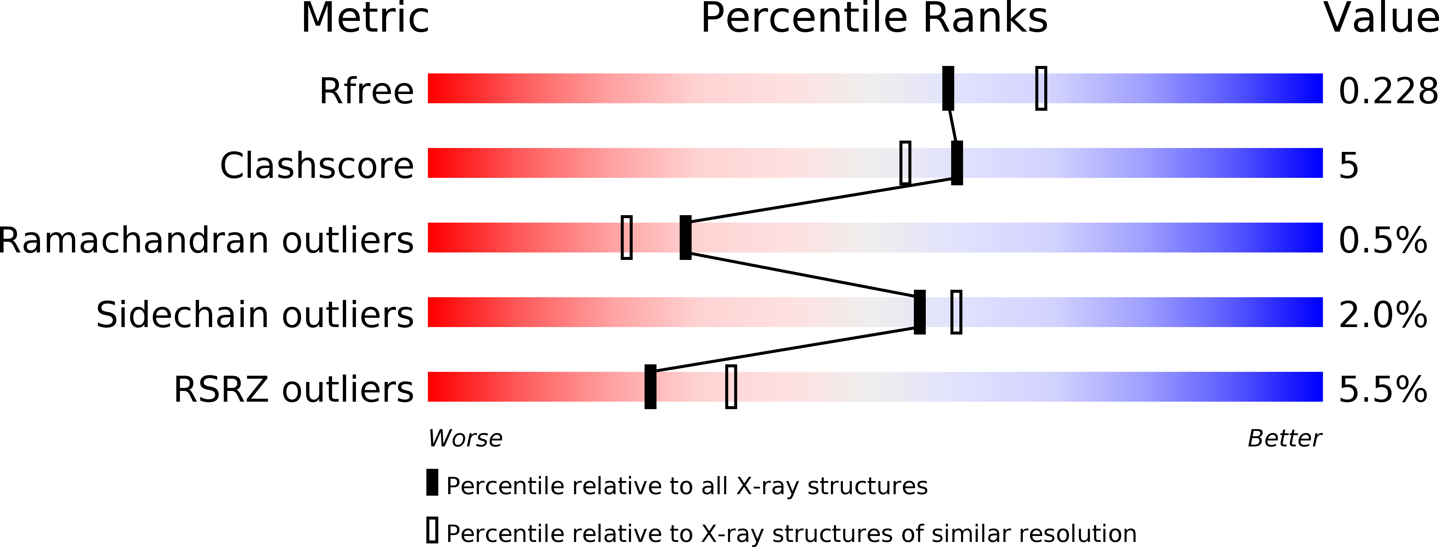
R-Value Free:
0.22
R-Value Work:
0.16
R-Value Observed:
0.17
Space Group:
C 1 2 1