Deposition Date
2016-09-24
Release Date
2017-01-11
Last Version Date
2024-03-06
Entry Detail
PDB ID:
5TF6
Keywords:
Title:
Structure and conformational plasticity of the U6 small nuclear ribonucleoprotein core
Biological Source:
Source Organism(s):
Saccharomyces cerevisiae (Taxon ID: 559292)
Saccharomyces cerevisiae (Taxon ID: 4932)
Saccharomyces cerevisiae (Taxon ID: 4932)
Expression System(s):
Method Details:
Experimental Method:
Resolution:
2.30 Å
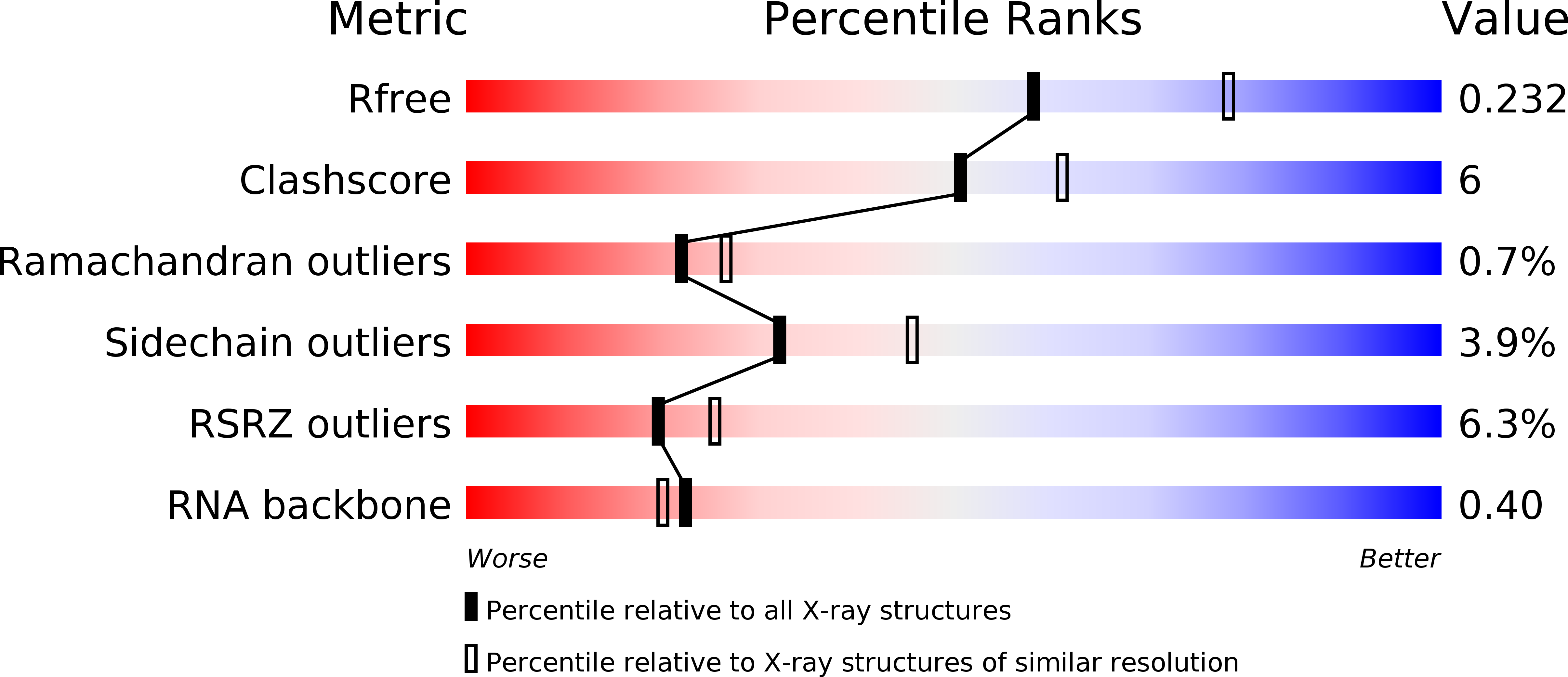
R-Value Free:
0.23
R-Value Work:
0.18
R-Value Observed:
0.18
Space Group:
P 21 21 21