5TDH image
Deposition Date 2016-09-19
Release Date 2016-11-09
Last Version Date 2024-03-20
Entry Detail
PDB ID:
5TDH
Keywords:
Title:
The crystal structure of the dominant negative mutant G protein alpha(i)-1-beta-1-gamma-2 G203A/A326S
Biological Source:
Source Organism:
Homo sapiens (Taxon ID: 9606)
Rattus norvegicus (Taxon ID: 10116)
Bos taurus (Taxon ID: 9913)
Host Organism:
Method Details:
Experimental Method:
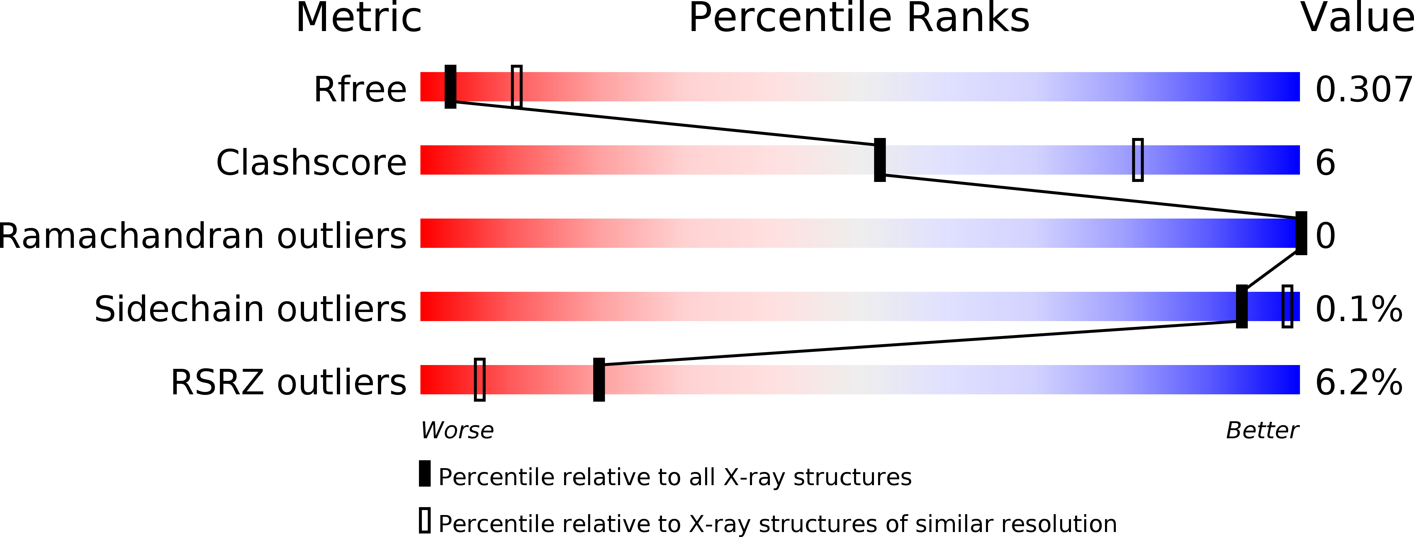
Resolution:
3.00 Å
R-Value Free:
0.30
R-Value Work:
0.27
R-Value Observed:
0.27
Space Group:
P 1 21 1
Macromolecular Entities
Structures with similar UniProt ID
Protein Blast
Polymer Type:polypeptide(L)
Molecule:Guanine nucleotide-binding protein G(i) subunit alpha-1
Gene (Uniprot):GNAI1
Mutagens:G203A,A326S
Chain IDs:A, D (auth: H)
Chain Length:354
Number of Molecules:2
Biological Source:Homo sapiens
Structures with similar UniProt ID
Protein Blast
Polymer Type:polypeptide(L)
Molecule:Guanine nucleotide-binding protein G(I)/G(S)/G(T) subunit beta-1
Gene (Uniprot):Gnb1
Chain IDs:B, E (auth: J)
Chain Length:342
Number of Molecules:2
Biological Source:Rattus norvegicus
Structures with similar UniProt ID
Protein Blast
Polymer Type:polypeptide(L)
Molecule:Guanine nucleotide-binding protein G(I)/G(S)/G(O) subunit gamma-2
Gene (Uniprot):GNG2
Chain IDs:C (auth: G), F (auth: K)
Chain Length:68
Number of Molecules:2
Biological Source:Bos taurus
Ligand Molecules
Primary Citation
The structural basis of the dominant negative phenotype of the G alpha i1 beta 1 gamma 2 G203A/A326S heterotrimer
Acta Pharmacol.Sin. 37 1259 1272 (2016)
PMID: 27498775 DOI: 10.1038/aps.2016.69

Abstact

AIM Dominant negative mutant G proteins have provided critical insight into the mechanisms of G protein-coupled receptor (GPCR) signaling, but the mechanisms underlying the dominant negative characteristics are not completely understood. The aim of this study was to determine the structure of the dominant negative Gαi1β1γ2 G203A/A326S complex (Gi-DN) and to reveal the structural basis of the mutation-induced phenotype of Gαi1β1γ2. METHODS The three subunits of the Gi-DN complex were co-expressed with a baculovirus expression system. The Gi-DN heterotrimer was purified, and the structure of its complex with GDP was determined through X-ray crystallography. RESULTS The Gi-DN heterotrimer structure revealed a dual mechanism underlying the dominant negative characteristics. The mutations weakened the hydrogen bonding network between GDP/GTP and the binding pocket residues, and increased the interactions in the Gα-Gβγ interface. Concomitantly, the Gi-DN heterotrimer adopted a conformation, in which the C-terminus of Gαi and the N-termini of both the Gβ and Gγ subunits were more similar to the GPCR-bound state compared with the wild type complex. From these structural observations, two additional mutations (T48F and D272F) were designed that completely abolish the GDP binding of the Gi-DN heterotrimer. CONCLUSION Overall, the results suggest that the mutations impede guanine nucleotide binding and Gα-Gβγ protein dissociation and favor the formation of the G protein/GPCR complex, thus blocking signal propagation. In addition, the structure provides a rationale for the design of other mutations that cause dominant negative effects in the G protein, as exemplified by the T48F and D272F mutations.

Legend

Protein

Chemical

Disease

Primary Citation of related structures