5TAR image
Deposition Date 2016-09-10
Release Date 2016-11-02
Last Version Date 2023-10-04
Entry Detail
PDB ID:
5TAR
Keywords:
Title:
Crystal structure of farnesylated and methylated kras4b in complex with PDE-delta (crystal form II - with ordered hypervariable region)
Biological Source:
Source Organism:
Homo sapiens (Taxon ID: 9606)
Method Details:
Experimental Method:
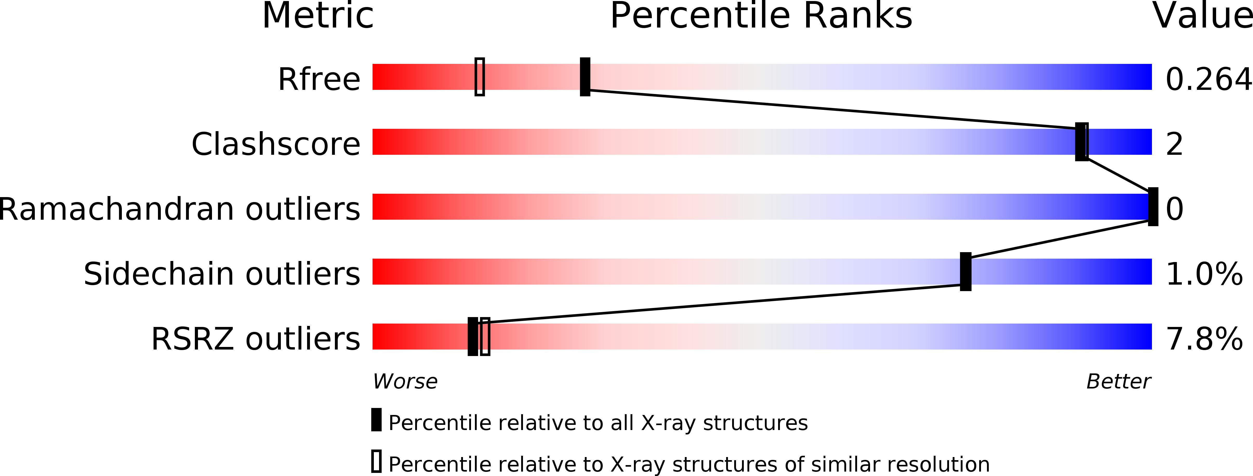
Resolution:
1.90 Å
R-Value Free:
0.26
R-Value Work:
0.22
R-Value Observed:
0.22
Space Group:
P 21 21 21
Macromolecular Entities
Polymer Type:polypeptide(L)
Molecule:GTPase KRas
Gene (Uniprot):KRAS
Chain IDs:A
Chain Length:185
Number of Molecules:1
Biological Source:Homo sapiens
Polymer Type:polypeptide(L)
Molecule:Retinal rod rhodopsin-sensitive cGMP 3',5'-cyclic phosphodiesterase subunit delta
Gene (Uniprot):PDE6D
Chain IDs:B
Chain Length:148
Number of Molecules:1
Biological Source:Homo sapiens
Modified Residue
Compound ID Chain ID Parent Comp ID Details 2D Image
CMT A CYS modified residue
Primary Citation
Structural basis of recognition of farnesylated and methylated KRAS4b by PDE delta.
Proc.Natl.Acad.Sci.USA 113 E6766 E6775 (2016)
PMID: 27791178 DOI: 10.1073/pnas.1615316113

Abstact

Farnesylation and carboxymethylation of KRAS4b (Kirsten rat sarcoma isoform 4b) are essential for its interaction with the plasma membrane where KRAS-mediated signaling events occur. Phosphodiesterase-δ (PDEδ) binds to KRAS4b and plays an important role in targeting it to cellular membranes. We solved structures of human farnesylated-methylated KRAS4b in complex with PDEδ in two different crystal forms. In these structures, the interaction is driven by the C-terminal amino acids together with the farnesylated and methylated C185 of KRAS4b that binds tightly in the central hydrophobic pocket present in PDEδ. In crystal form II, we see the full-length structure of farnesylated-methylated KRAS4b, including the hypervariable region. Crystal form I reveals structural details of farnesylated-methylated KRAS4b binding to PDEδ, and crystal form II suggests the potential binding mode of geranylgeranylated-methylated KRAS4b to PDEδ. We identified a 5-aa-long sequence motif (Lys-Ser-Lys-Thr-Lys) in KRAS4b that may enable PDEδ to bind both forms of prenylated KRAS4b. Structure and sequence analysis of various prenylated proteins that have been previously tested for binding to PDEδ provides a rationale for why some prenylated proteins, such as KRAS4a, RalA, RalB, and Rac1, do not bind to PDEδ. Comparison of all four available structures of PDEδ complexed with various prenylated proteins/peptides shows the presence of additional interactions due to a larger protein-protein interaction interface in KRAS4b-PDEδ complex. This interface might be exploited for designing an inhibitor with minimal off-target effects.

Legend

Protein

Chemical

Disease

Primary Citation of related structures