Deposition Date
2016-09-09
Release Date
2016-11-02
Last Version Date
2023-10-04
Entry Detail
PDB ID:
5T9C
Keywords:
Title:
Crystal structure of B. subtilis 168 GlpQ in complex with glycerol-3-phosphate (1 hour soak)
Biological Source:
Source Organism(s):
Bacillus subtilis (Taxon ID: 224308)
Expression System(s):
Method Details:
Experimental Method:
Resolution:
1.48 Å
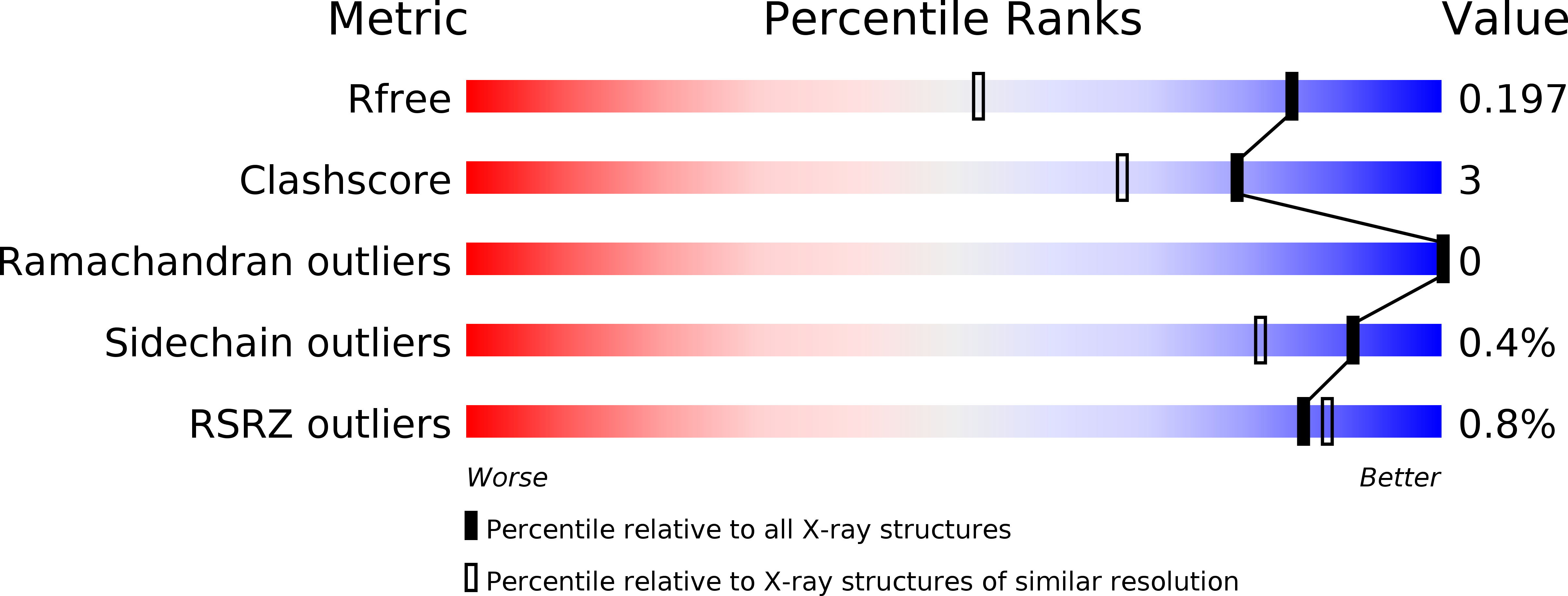
R-Value Free:
0.19
R-Value Work:
0.16
R-Value Observed:
0.17
Space Group:
P 21 21 21