Deposition Date
2016-09-08
Release Date
2017-04-05
Last Version Date
2024-01-17
Entry Detail
PDB ID:
5T8R
Keywords:
Title:
Crystal structure of human BAZ2A PHD zinc finger in complex with unmodified H3 10-mer
Biological Source:
Source Organism(s):
Homo sapiens (Taxon ID: 9606)
Expression System(s):
Method Details:
Experimental Method:
Resolution:
2.40 Å
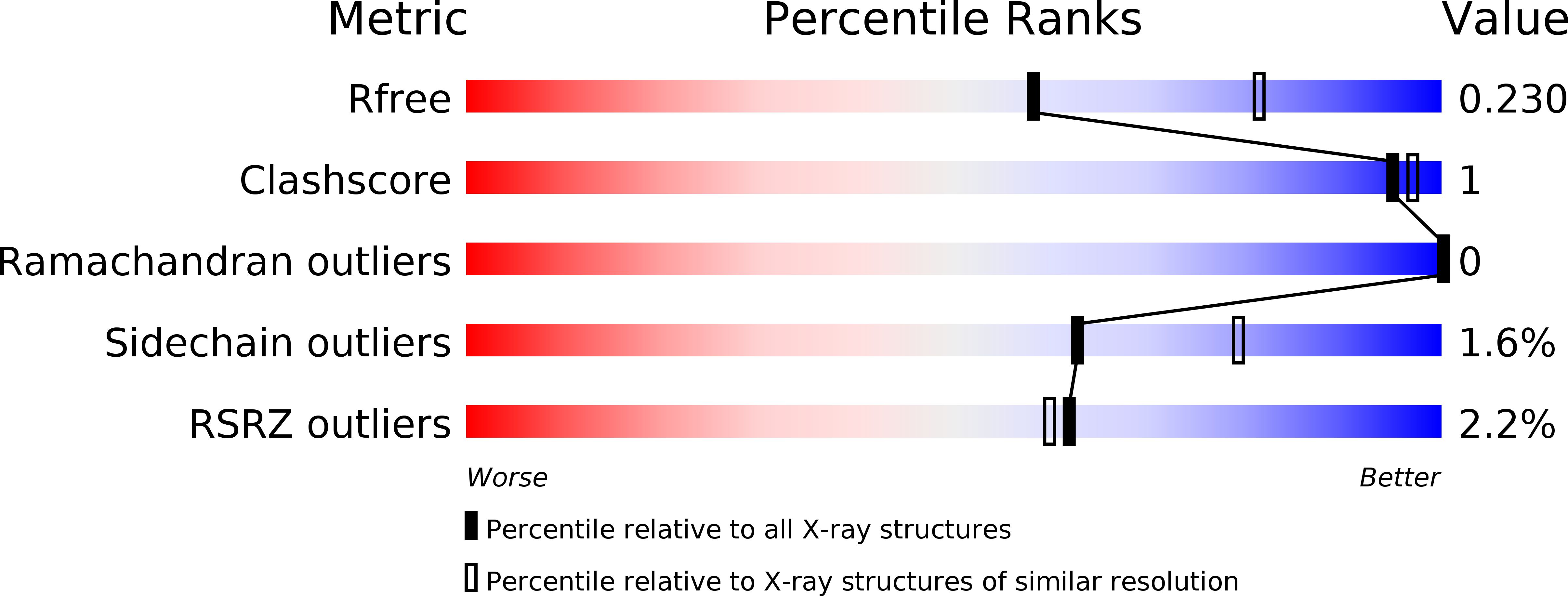
R-Value Free:
0.23
R-Value Work:
0.18
R-Value Observed:
0.18
Space Group:
P 43 21 2