
Deposition Date
2016-09-07
Release Date
2017-03-01
Last Version Date
2024-04-03
Entry Detail
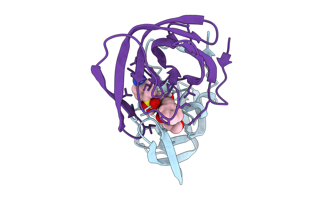
PDB ID:
5T8H
Keywords:
Title:
Joint X-ray/neutron structure of HIV-1 protease triple mutant (V32I,I47V,V82I) with amprenavir at pH 6.0
Biological Source:
Source Organism:
Human immunodeficiency virus 1 (Taxon ID: 11676)
Host Organism:
Method Details:
Experimental Method:
R-Value Free:
['0.21
R-Value Work:
['0.19
R-Value Observed:
['?', '?'].00
Space Group:
P 21 21 2
