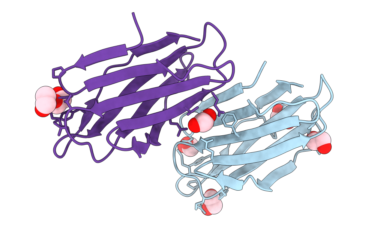
5T7A image
Deposition Date 2016-09-02
Release Date 2017-08-23
Last Version Date 2024-03-06
Entry Detail
PDB ID:
5T7A
Title:
Crystal structure of Br derivative BhCBM56
Biological Source:
Method Details:
Experimental Method:
Resolution:
1.60 Å
R-Value Free:
0.17
R-Value Work:
0.14
R-Value Observed:
0.14
Space Group:
C 1 2 1
Macromolecular Entities
Polymer Type:polypeptide(L)
Molecule:BH0236 protein
Gene (Uniprot):BH0236
Chain IDs:A, B
Chain Length:118
Number of Molecules:2
Biological Source:Bacillus halodurans (strain ATCC BAA-125 / DSM 18197 / FERM 7344 / JCM 9153 / C-125)
Primary Citation
Properties of a family 56 carbohydrate-binding module and its role in the recognition and hydrolysis of beta-1,3-glucan.
J. Biol. Chem. 292 16955 16968 (2017)
PMID: 28827308 DOI: 10.1074/jbc.M117.806711

Abstact

BH0236 from Bacillus halodurans is a multimodular β-1,3-glucanase comprising an N-terminal family 81 glycoside hydrolase catalytic module, an internal family 6 carbohydrate-binding module (CBM) that binds the nonreducing end of β-1,3-glucan chains, and an uncharacterized C-terminal module classified into CBM family 56. Here, we determined that this latter CBM, BhCBM56, bound the soluble β-1,3-glucan laminarin with a dissociation constant (Kd) of ∼26 μm and displayed higher affinity for insoluble β-1,3-glucans with Kd values of ∼2-10 μm but lacked affinity for β-1,3-glucooligosaccharides. The X-ray crystal structure of BhCBM56 and NMR-derived chemical shift mapping of the binding site revealed a β-sandwich fold, with the face of one β-sheet possessing the β-1,3-glucan-binding surface. On the basis of the functional and structural properties of BhCBM56, we propose that it binds a quaternary polysaccharide structure, most likely the triple helix adopted by polymerized β-1,3-glucans. Consistent with the BhCBM56 and BhCBM6/56 binding profiles, deletion of the CBM56 from BH0236 decreased activity of the enzyme on the insoluble β-1,3-glucan curdlan but not on soluble laminarin; additional deletion of the CBM6 also did not affect laminarin degradation but further decreased curdlan hydrolysis. The pseudo-atomic solution structure of BH0236 determined by small-angle X-ray scattering revealed structural insights into the nature of avid binding by the BhCBM6/56 pair and how the orientation of the active site in the catalytic module factors into recognition and degradation of β-1,3-glucans. Our findings reinforce the notion that catalytic modules and their cognate CBMs have complementary specificities, including targeting of polysaccharide quaternary structure.

Legend

Protein

Chemical

Disease

Primary Citation of related structures