Deposition Date
2016-09-01
Release Date
2017-01-25
Last Version Date
2024-01-17
Entry Detail
PDB ID:
5T69
Keywords:
Title:
The HhoA protease from Synechocystis sp. PCC 6803, active site mutant
Biological Source:
Source Organism(s):
Synechocystis sp. PCC 6803 substr. Kazusa (Taxon ID: 1111708)
Expression System(s):
Method Details:
Experimental Method:
Resolution:
2.10 Å
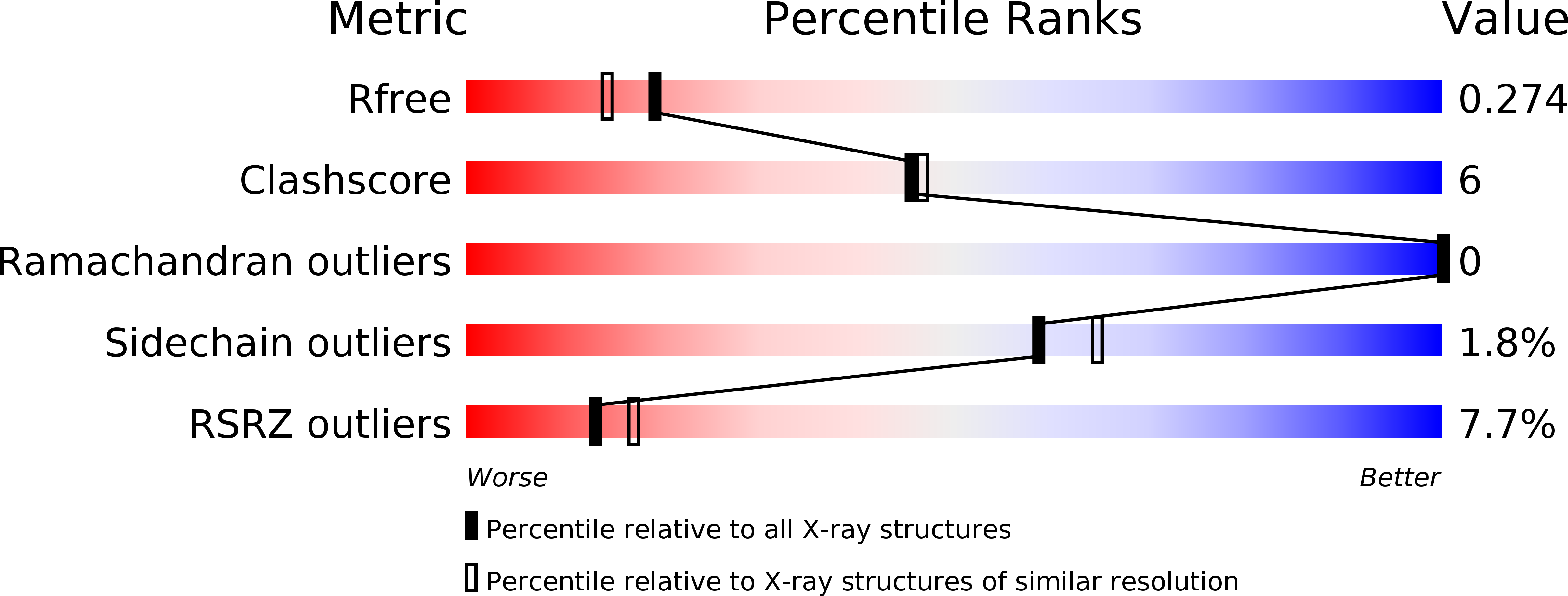
R-Value Free:
0.27
R-Value Work:
0.24
R-Value Observed:
0.24
Space Group:
H 3 2