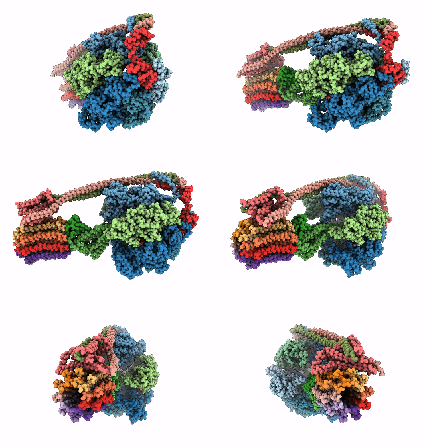
5T4Q image
Entry Detail
PDB ID:
5T4Q
EMDB ID:
Keywords:
Title:
Autoinhibited E. coli ATP synthase state 3
Biological Source:
Source Organism:
Host Organism:
PDB Version:
Deposition Date:
2016-08-29
Release Date:
2017-01-04
Method Details:
Experimental Method:
Resolution:
8.53 Å
Aggregation State:
PARTICLE
Reconstruction Method:
SINGLE PARTICLE
Macromolecular Entities
Polymer Type:polypeptide(L)
Description:ATP synthase subunit alpha
Chain IDs:A, B, C
Chain Length:513
Number of Molecules:3
Biological Source:Escherichia coli
Polymer Type:polypeptide(L)
Description:ATP synthase subunit beta
Chain IDs:D, E, F
Chain Length:471
Number of Molecules:3
Biological Source:Escherichia coli
Polymer Type:polypeptide(L)
Description:ATP synthase gamma chain
Chain IDs:G
Chain Length:287
Number of Molecules:1
Biological Source:Escherichia coli
Polymer Type:polypeptide(L)
Description:ATP synthase epsilon chain
Chain IDs:H
Chain Length:139
Number of Molecules:1
Biological Source:Escherichia coli
Polymer Type:polypeptide(L)
Description:ATP synthase subunit b
Chain IDs:I, J
Chain Length:155
Number of Molecules:2
Biological Source:Escherichia coli
Polymer Type:polypeptide(L)
Description:ATP synthase subunit a
Chain IDs:K
Chain Length:271
Number of Molecules:1
Biological Source:Escherichia coli
Polymer Type:polypeptide(L)
Description:ATP synthase subunit delta
Chain IDs:L
Chain Length:177
Number of Molecules:1
Biological Source:Escherichia coli
Polymer Type:polypeptide(L)
Description:ATP synthase subunit c
Chain IDs:M, N, O, P, Q, R, S, T, U, V
Chain Length:79
Number of Molecules:10
Biological Source:Escherichia coli
Primary Citation
Cryo-EM structures of the autoinhibitedE. coliATP synthase in three rotational states.
Elife 5 ? ? (2016)
PMID: 28001127 DOI: 10.7554/eLife.21598

Abstact

A molecular model that provides a framework for interpreting the wealth of functional information obtained on the E. coli F-ATP synthase has been generated using cryo-electron microscopy. Three different states that relate to rotation of the enzyme were observed, with the central stalk's ε subunit in an extended autoinhibitory conformation in all three states. The Fo motor comprises of seven transmembrane helices and a decameric c-ring and invaginations on either side of the membrane indicate the entry and exit channels for protons. The proton translocating subunit contains near parallel helices inclined by ~30° to the membrane, a feature now synonymous with rotary ATPases. For the first time in this rotary ATPase subtype, the peripheral stalk is resolved over its entire length of the complex, revealing the F1 attachment points and a coiled-coil that bifurcates toward the membrane with its helices separating to embrace subunit a from two sides.

Legend

Protein

Chemical

Disease

Primary Citation of related structures