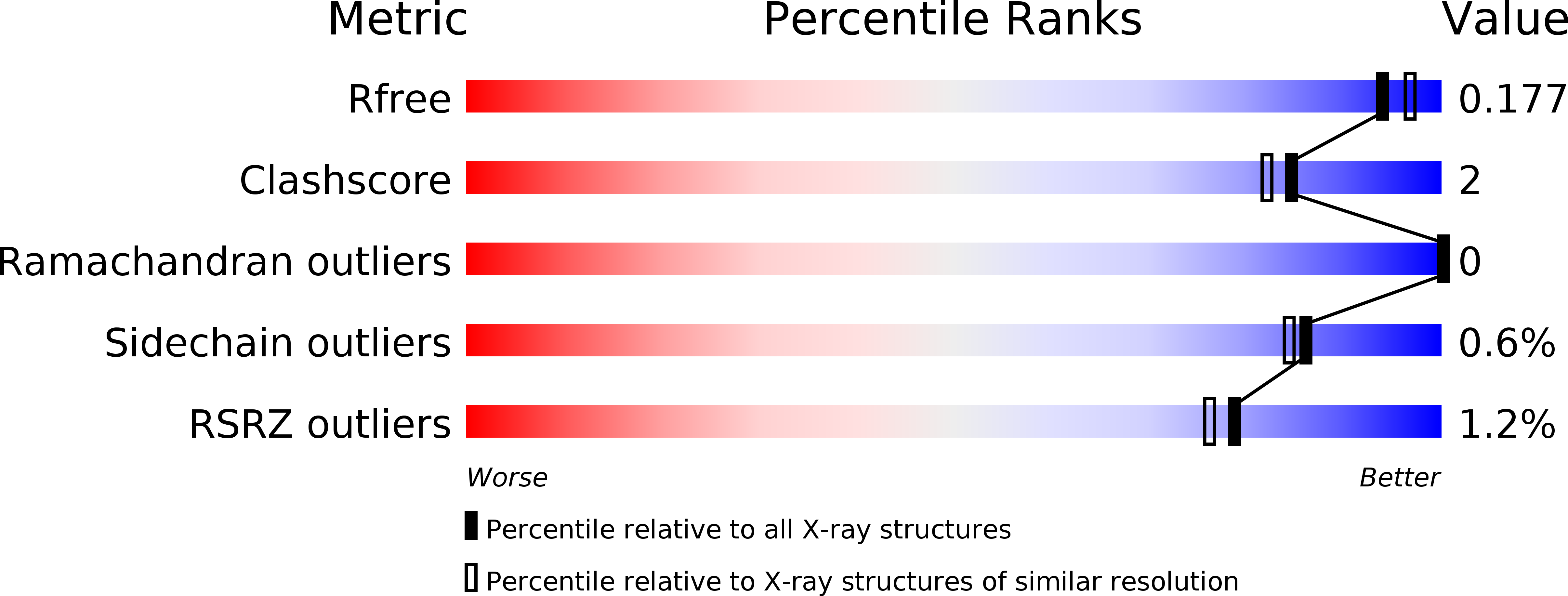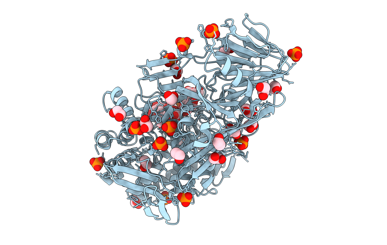
5T4G image
Deposition Date 2016-08-29
Release Date 2017-06-28
Last Version Date 2023-10-04
Entry Detail
PDB ID:
5T4G
Keywords:
Title:
Crystal structure of BhGH81 in complex with laminarin
Biological Source:
Source Organism:
Host Organism:
Method Details:
Experimental Method:
Resolution:
1.80 Å
R-Value Free:
0.17
R-Value Work:
0.16
R-Value Observed:
0.16
Space Group:
P 21 21 21
Macromolecular Entities
Structures with similar UniProt ID
Protein Blast
Polymer Type:polypeptide(L)
Molecule:Glycoside Hydrolase
Gene (Uniprot):BH0236
Chain IDs:A
Chain Length:783
Number of Molecules:1
Biological Source:Bacillus halodurans
Peptide-like Molecules
PRD_900024
Primary Citation
Structural Analysis of a Family 81 Glycoside Hydrolase Implicates Its Recognition of beta-1,3-Glucan Quaternary Structure.
Structure 25 1348 1359.e3 (2017)
PMID: 28781080 DOI: 10.1016/j.str.2017.06.019

Abstact

Family 81 glycoside hydrolases (GHs), which are known to cleave β-1,3-glucans, are found in archaea, bacteria, eukaryotes, and viruses. Here we examine the structural and functional features of the GH81 catalytic module, BhGH81, from the Bacillus halodurans protein BH0236 to probe the molecular basis of β-1,3-glucan recognition and cleavage. BhGH81 displayed activity on laminarin, curdlan, and pachyman, but not scleroglucan; the enzyme also cleaved β-1,3-glucooligosaccharides as small as β-1,3-glucotriose. The crystal structures of BhGH81 in complex with various β-1,3-glucooligosaccharides revealed distorted sugars in the -1 catalytic subsite and an arrangement consistent with an inverting catalytic mechanism having a proposed conformational itinerary of 2S0 → 2,5B‡ → 5S1. Notably, the architecture of the catalytic site, location of an adjacent ancillary β-1,3-glucan binding site, and the surface properties of the enzyme indicate the likely ability to recognize the double and/or triple-helical quaternary structures adopted by β-1,3-glucans.

Legend

Protein

Chemical

Disease

Primary Citation of related structures