5T4D image
Entry Detail
PDB ID:
5T4D
EMDB ID:
Keywords:
Title:
Cryo-EM structure of Polycystic Kidney Disease protein 2 (PKD2), residues 198-703
Biological Source:
Source Organism:
Host Organism:
PDB Version:
Deposition Date:
2016-08-29
Release Date:
2016-11-02
Method Details:
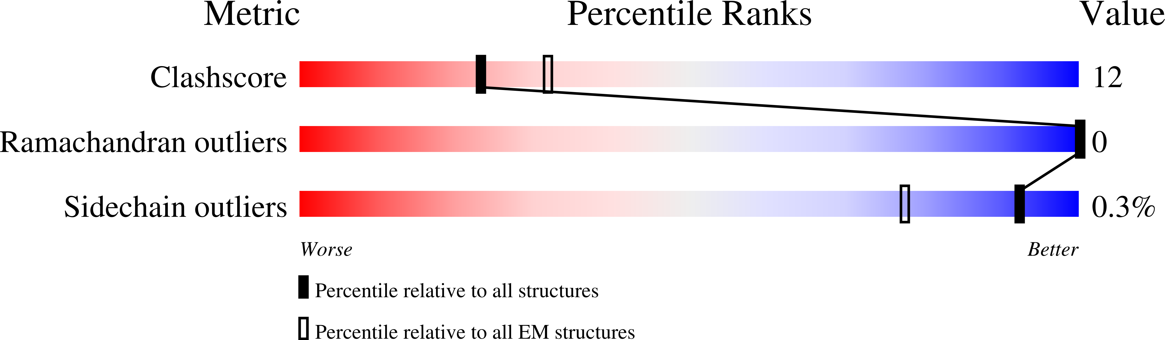
Experimental Method:
Resolution:
3.00 Å
Aggregation State:
PARTICLE
Reconstruction Method:
SINGLE PARTICLE
Macromolecular Entities
Polymer Type:polypeptide(L)
Description:hPKD:198-703, Polycystin-2
Chain IDs:A, B, C, D
Chain Length:510
Number of Molecules:4
Biological Source:Homo sapiens
Ligand Molecules
Primary Citation
The Structure of the Polycystic Kidney Disease Channel PKD2 in Lipid Nanodiscs.
Cell 167 763 773.e11 (2016)
PMID: 27768895 DOI: 10.1016/j.cell.2016.09.048

Abstact

The Polycystic Kidney Disease 2 (Pkd2) gene is mutated in autosomal dominant polycystic kidney disease (ADPKD), one of the most common human monogenic disorders. Here, we present the cryo-EM structure of PKD2 in lipid bilayers at 3.0 Å resolution, which establishes PKD2 as a homotetrameric ion channel and provides insight into potential mechanisms for its activation. The PKD2 voltage-sensor domain retains two of four gating charges commonly found in those of voltage-gated ion channels. The PKD2 ion permeation pathway is constricted at the selectivity filter and near the cytoplasmic end of S6, suggesting that two gates regulate ion conduction. The extracellular domain of PKD2, a hotspot for ADPKD pathogenic mutations, contributes to channel assembly and strategically interacts with the transmembrane core, likely serving as a physical substrate for extracellular stimuli to allosterically gate the channel. Finally, our structure establishes the molecular basis for the majority of pathogenic mutations in Pkd2-related ADPKD.

Legend

Protein

Chemical

Disease

Primary Citation of related structures