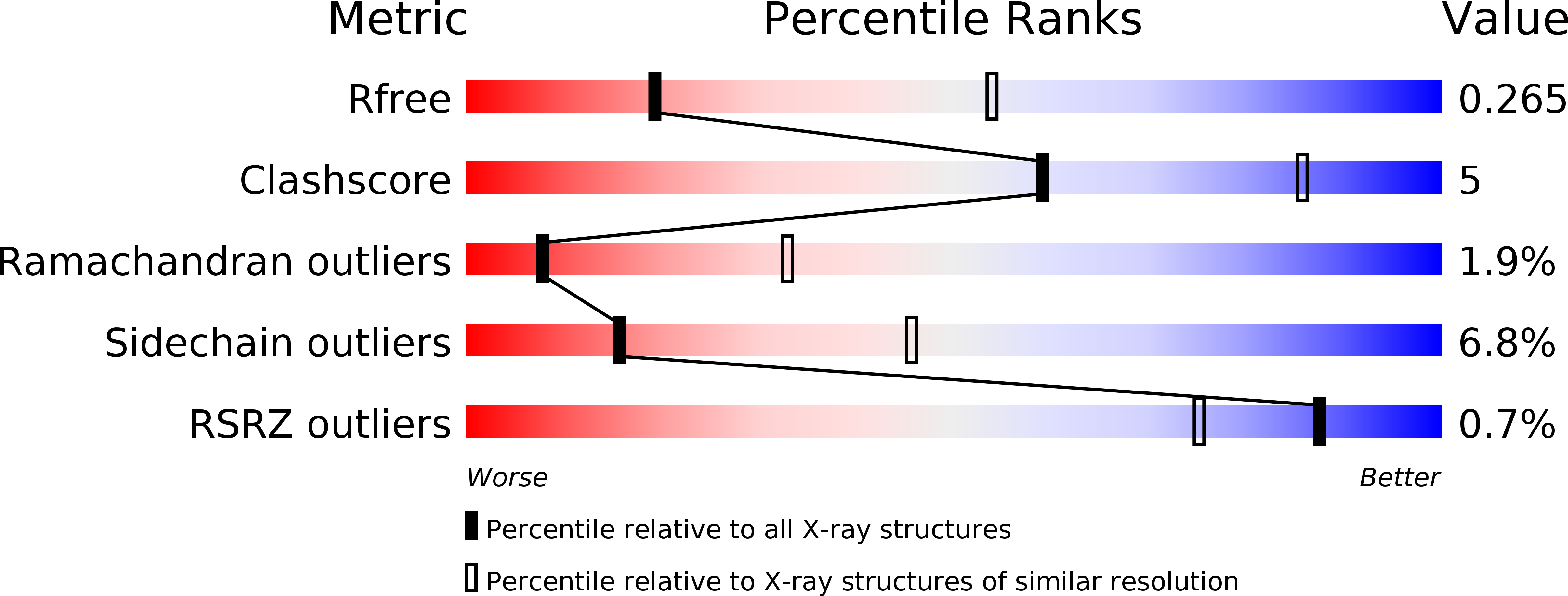
5T1D image
Deposition Date 2016-08-18
Release Date 2016-12-28
Last Version Date 2024-10-16
Entry Detail
PDB ID:
5T1D
Keywords:
Title:
Crystal structure of EBV gHgL/gp42/E1D1 complex
Biological Source:
Method Details:
Experimental Method:
Resolution:
3.10 Å
R-Value Free:
0.26
R-Value Work:
0.21
R-Value Observed:
0.21
Space Group:
I 2 2 2
Macromolecular Entities
Structures with similar UniProt ID
Protein Blast
Polymer Type:polypeptide(L)
Molecule:Envelope glycoprotein H
Gene (Uniprot):gH
Chain IDs:A
Chain Length:655
Number of Molecules:1
Biological Source:Epstein-Barr virus (strain B95-8)
Structures with similar UniProt ID
Protein Blast
Polymer Type:polypeptide(L)
Molecule:Envelope glycoprotein L
Gene (Uniprot):gL
Chain IDs:B
Chain Length:112
Number of Molecules:1
Biological Source:Epstein-Barr virus (strain B95-8)
Structures with similar UniProt ID
Protein Blast
Polymer Type:polypeptide(L)
Molecule:Glycoprotein 42
Gene (Uniprot):BZLF2
Chain IDs:C
Chain Length:191
Number of Molecules:1
Biological Source:Epstein-Barr virus (strain GD1)
Polymer Type:polypeptide(L)
Molecule:E1D1 IgG2a heavy chain
Chain IDs:D (auth: H)
Chain Length:213
Number of Molecules:1
Biological Source:Mus musculus
Polymer Type:polypeptide(L)
Molecule:E1D1 IgG2a light chain
Chain IDs:E (auth: L)
Chain Length:217
Number of Molecules:1
Biological Source:Mus musculus
Ligand Molecules
Primary Citation
Structural basis for Epstein-Barr virus host cell tropism mediated by gp42 and gHgL entry glycoproteins.
Nat Commun 7 13557 13557 (2016)
PMID: 27929061 DOI: 10.1038/ncomms13557

Abstact

Herpesvirus entry into host cells is mediated by multiple virally encoded receptor binding and membrane fusion glycoproteins. Despite their importance in host cell tropism and associated disease pathology, the underlying and essential interactions between these viral glycoproteins remain poorly understood. For Epstein-Barr virus (EBV), gHgL/gp42 complexes bind HLA class II to activate membrane fusion with B cells, but gp42 inhibits fusion and entry into epithelial cells. To clarify the mechanism by which gp42 controls the cell specificity of EBV infection, here we determined the structure of gHgL/gp42 complex bound to an anti-gHgL antibody (E1D1). The critical regulator of EBV tropism is the gp42 N-terminal domain, which tethers the HLA-binding domain to gHgL by wrapping around the exterior of three gH domains. Both the gp42 N-terminal domain and E1D1 selectively inhibit epithelial-cell fusion; however, they engage distinct surfaces of gHgL. These observations clarify key determinants of EBV host cell tropism.

Legend

Protein

Chemical

Disease

Primary Citation of related structures