Deposition Date
2016-08-14
Release Date
2016-10-19
Last Version Date
2024-10-23
Entry Detail
PDB ID:
5SZR
Keywords:
Title:
Protocadherin Gamma B2 extracellular cadherin domains 3-6
Biological Source:
Source Organism(s):
Mus musculus (Taxon ID: 10090)
Expression System(s):
Method Details:
Experimental Method:
Resolution:
2.30 Å
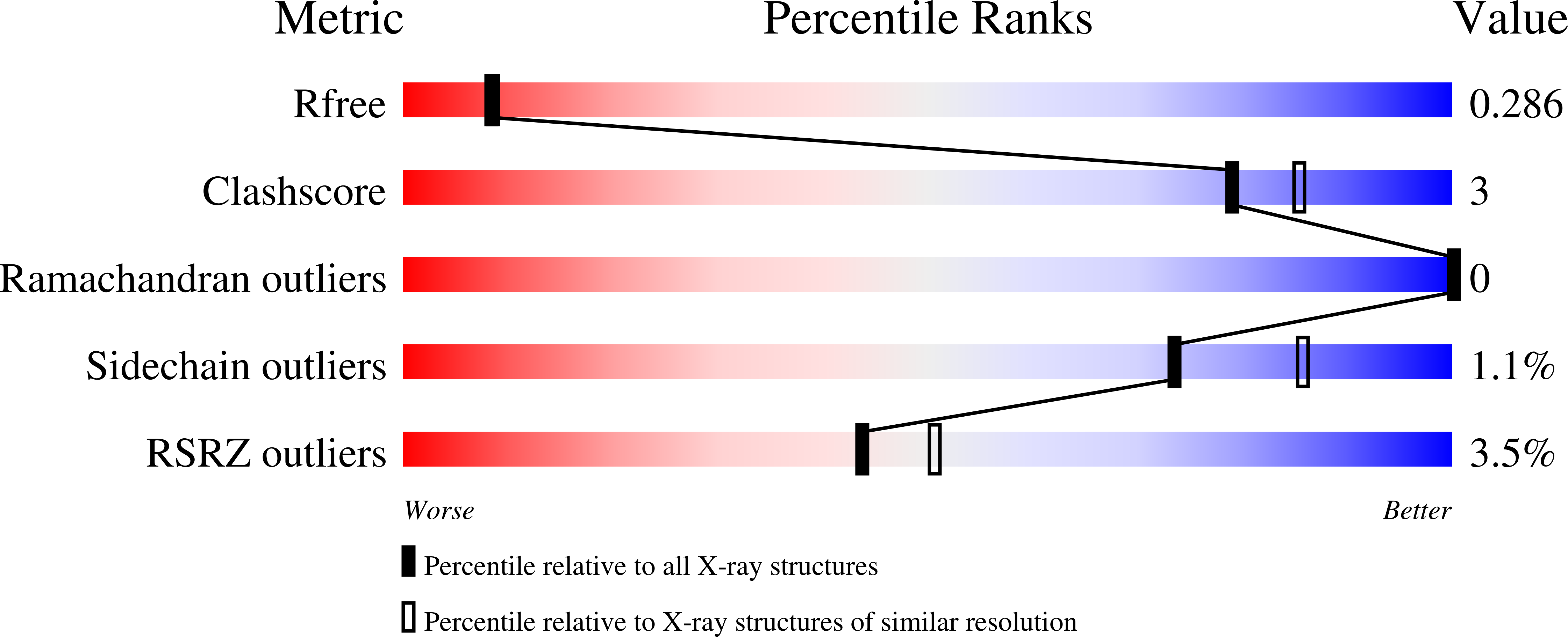
R-Value Free:
0.27
R-Value Work:
0.24
R-Value Observed:
0.25
Space Group:
P 41 21 2