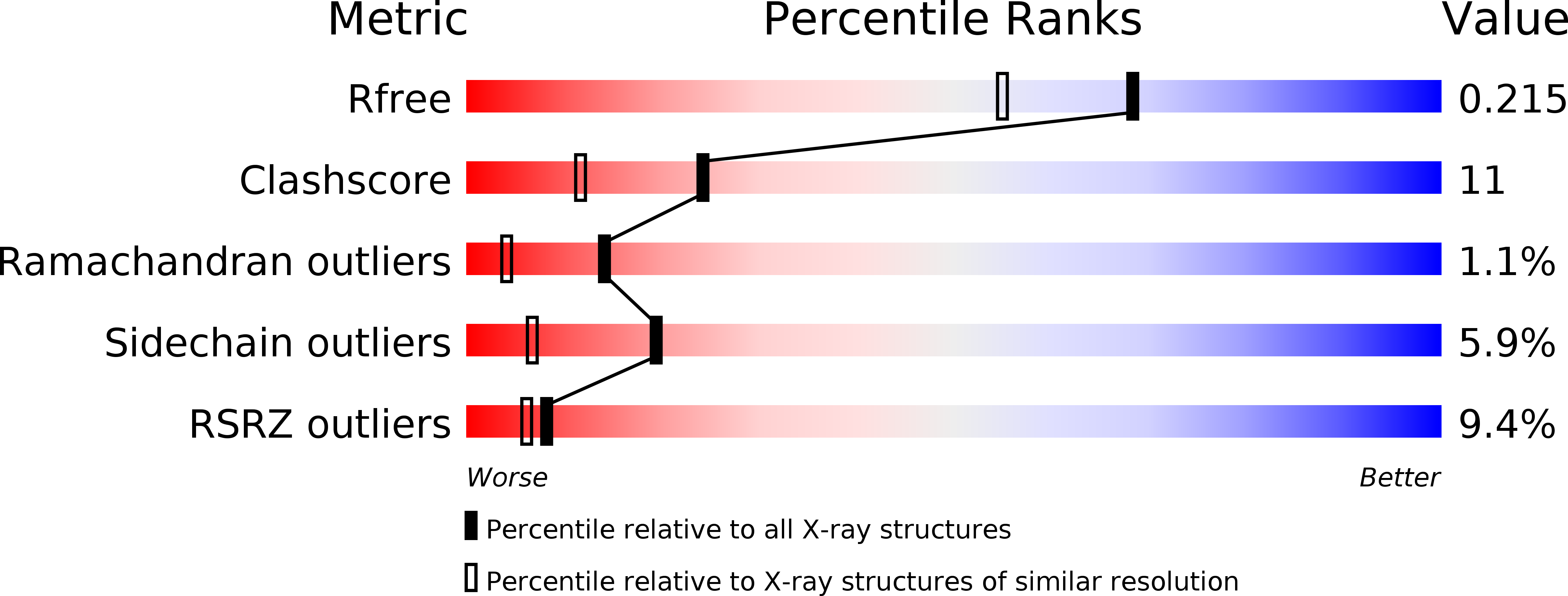
5SYB image
Entry Detail
PDB ID:
5SYB
Keywords:
Title:
Crystal structure of human PHF5A
Biological Source:
Source Organism:
Host Organism:
PDB Version:
Deposition Date:
2016-08-10
Release Date:
2016-09-07
Method Details:
Experimental Method:
Resolution:
1.82 Å
R-Value Free:
0.20
R-Value Work:
0.16
R-Value Observed:
0.16
Space Group:
P 21 3
Macromolecular Entities
Polymer Type:polypeptide(L)
Description:PHD finger-like domain-containing protein 5A
Mutations:C40S
Chain IDs:A, B
Chain Length:113
Number of Molecules:2
Biological Source:Homo sapiens
Primary Citation
Splicing modulators act at the branch point adenosine binding pocket defined by the PHF5A-SF3b complex.
Nat Commun 8 15522 15522 (2017)
PMID: 28541300 DOI: 10.1038/ncomms15522

Abstact

Pladienolide, herboxidiene and spliceostatin have been identified as splicing modulators that target SF3B1 in the SF3b subcomplex. Here we report that PHF5A, another component of this subcomplex, is also targeted by these compounds. Mutations in PHF5A-Y36, SF3B1-K1071, SF3B1-R1074 and SF3B1-V1078 confer resistance to these modulators, suggesting a common interaction site. RNA-seq analysis reveals that PHF5A-Y36C has minimal effect on basal splicing but inhibits the global action of splicing modulators. Moreover, PHF5A-Y36C alters splicing modulator-induced intron-retention/exon-skipping profile, which correlates with the differential GC content between adjacent introns and exons. We determine the crystal structure of human PHF5A demonstrating that Y36 is located on a highly conserved surface. Analysis of the cryo-EM spliceosome Bact complex shows that the resistance mutations cluster in a pocket surrounding the branch point adenosine, suggesting a competitive mode of action. Collectively, we propose that PHF5A-SF3B1 forms a central node for binding to these splicing modulators.

Legend

Protein

Chemical

Disease

Primary Citation of related structures