
Deposition Date
2016-08-10
Release Date
2016-08-31
Last Version Date
2024-11-06
Entry Detail
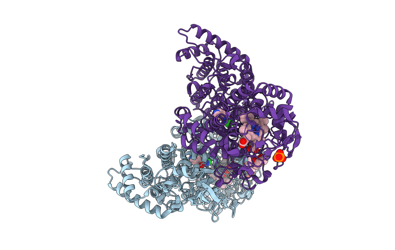
PDB ID:
5SXQ
Keywords:
Title:
Crystal structure of B. pseudomallei KatG with isonicotinic acid hydrazide bound
Biological Source:
Source Organism(s):
Burkholderia pseudomallei (strain 1710b) (Taxon ID: 320372)
Expression System(s):
Method Details:
Experimental Method:
Resolution:
2.10 Å
R-Value Free:
0.18
R-Value Work:
0.15
R-Value Observed:
0.15
Space Group:
P 21 21 21
