5SX5 image
Entry Detail
PDB ID:
5SX5
Title:
Crystal Structure of panitumumab in complex with epidermal growth factor receptor domain 3 mutant S468R.
Biological Source:
Source Organism:
Host Organism:
PDB Version:
Deposition Date:
2016-08-09
Release Date:
2016-10-05
Method Details:
Experimental Method:
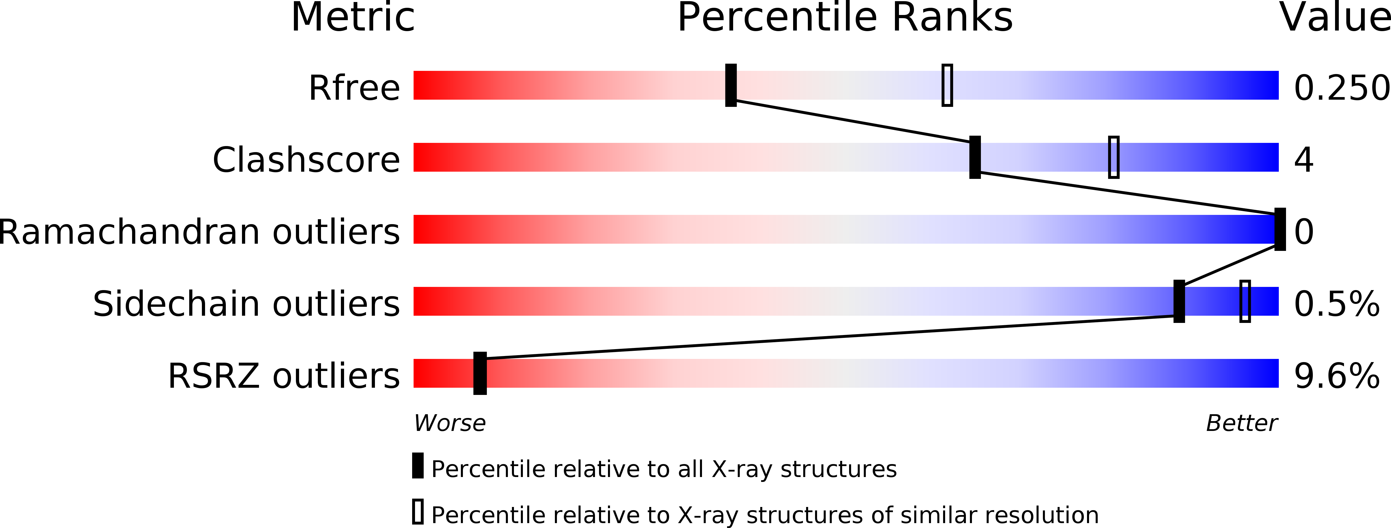
Resolution:
2.50 Å
R-Value Free:
0.24
R-Value Work:
0.22
R-Value Observed:
0.22
Space Group:
P 21 21 21
Macromolecular Entities
Polymer Type:polypeptide(L)
Description:Panitumumab Fab Heavy Chain
Chain IDs:B (auth: J), D (auth: H)
Chain Length:221
Number of Molecules:2
Biological Source:Homo sapiens
Polymer Type:polypeptide(L)
Description:Panitumumab Fab Light Chain
Chain IDs:A (auth: K), C (auth: L)
Chain Length:214
Number of Molecules:2
Biological Source:Homo sapiens
Polymer Type:polypeptide(L)
Description:Epidermal growth factor receptor
Chain IDs:E (auth: M), F (auth: N)
Chain Length:201
Number of Molecules:2
Biological Source:Homo sapiens
Primary Citation
The Panitumumab EGFR Complex Reveals a Binding Mechanism That Overcomes Cetuximab Induced Resistance.
Plos One 11 e0163366 e0163366 (2016)
PMID: 27658254 DOI: 10.1371/journal.pone.0163366

Abstact

Panitumumab and cetuximab target the epidermal growth factor receptor for the treatment of metastatic colorectal cancer. These therapies provide a significant survival benefit to patients with metastatic colorectal cancer with wild-type RAS. A single point mutation in the ectodomain of EGFR (S468R) confers acquired or secondary resistance in cetuximab treated patients, which is not observed in panitumumab-treated patients. Structural and biophysical studies presented here show this mutation directly blocks cetuximab binding to EGFR domain III and describes a unique mechanism by which panitumumab uses a central cavity to accommodate this mutation.

Legend

Protein

Chemical

Disease

Primary Citation of related structures