
Deposition Date
2016-08-08
Release Date
2017-02-15
Last Version Date
2024-10-16
Entry Detail
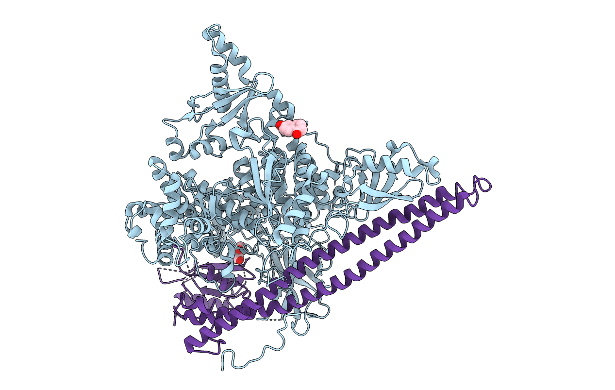
PDB ID:
5SWR
Keywords:
Title:
Crystal Structure of PI3Kalpha in complex with fragments 20 and 26
Biological Source:
Source Organism(s):
Homo sapiens (Taxon ID: 9606)
Expression System(s):
Method Details:
Experimental Method:
Resolution:
3.31 Å
R-Value Free:
0.27
R-Value Work:
0.19
R-Value Observed:
0.20
Space Group:
P 21 21 21
