5SV6 image
Deposition Date 2016-08-04
Release Date 2017-07-12
Last Version Date 2024-11-20
Entry Detail
PDB ID:
5SV6
Title:
Crystal structure of MxaJ from Methlophaga aminisulfidivorans MPT
Biological Source:
Source Organism:
Method Details:
Experimental Method:
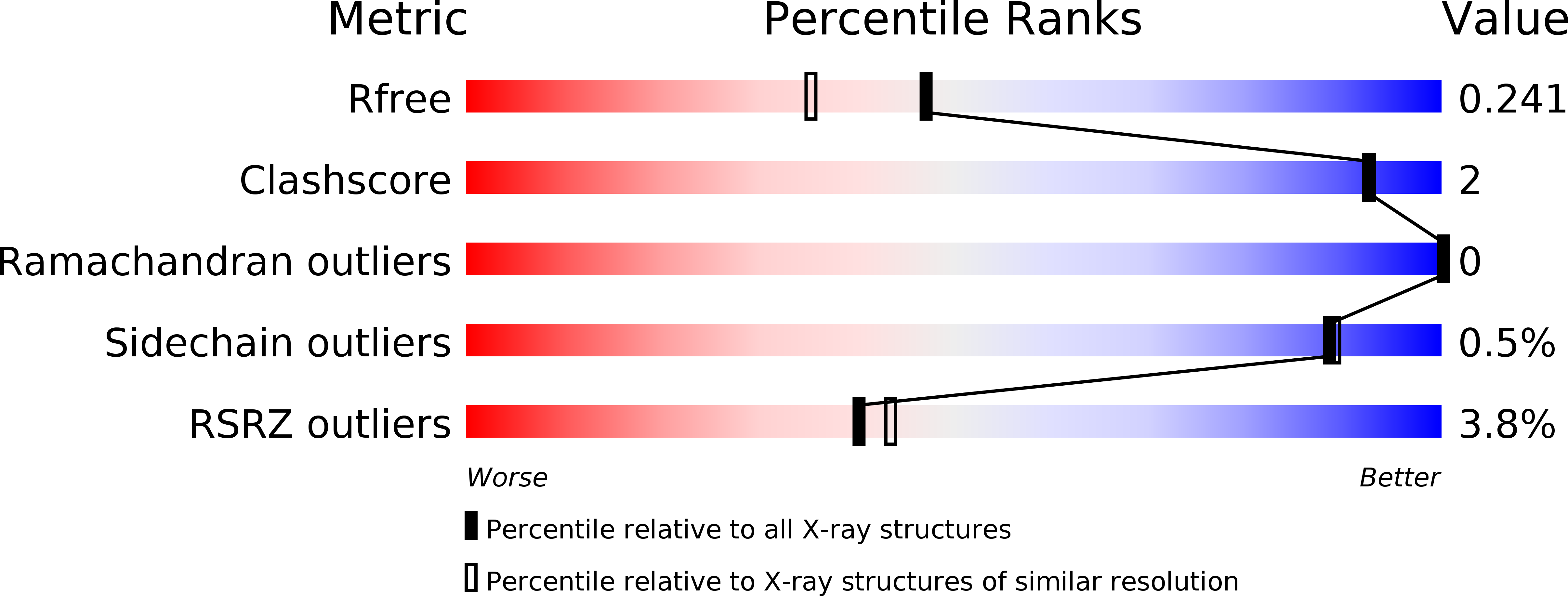
Resolution:
1.92 Å
R-Value Free:
0.23
R-Value Work:
0.17
R-Value Observed:
0.18
Space Group:
P 21 21 21
Macromolecular Entities
Structures with similar UniProt ID
Protein Blast
Polymer Type:polypeptide(L)
Molecule:Extracellular solute-binding protein, family 3
Gene (Uniprot):mxaJ
Chain IDs:A
Chain Length:291
Number of Molecules:1
Biological Source:Methylophaga aminisulfidivorans MP
Modified Residue
Compound ID Chain ID Parent Comp ID Details 2D Image
MSE A MET modified residue
Ligand Molecules
Primary Citation
MxaJ structure reveals a periplasmic binding protein-like architecture with unique secondary structural elements
Proteins 85 1379 1386 (2017)
PMID: 28295618 DOI: 10.1002/prot.25283

Abstact

MxaJ is a component of type II methanol dehydrogenase (MDH) that mediates electron transfer during methanol oxidation in methanotrophic bacteria. However, little is known about how MxaJ structurally cooperates with MDH and Cytochrome cL . Here, we report for the first time the crystal structure of MxaJ. MxaJ consists of eight α-helices and six β-strands, and resembles the "bi-lobate" folding architecture found in periplasmic binding proteins. Distinctive features of MxaJ include prominent loops and a β-strand around the hinge region supporting the ligand-binding cavity, which might provide a more favorable framework for interacting with proteins rather than small molecules. Proteins 2017; 85:1379-1386. © 2017 Wiley Periodicals, Inc.

Legend

Protein

Chemical

Disease

Primary Citation of related structures