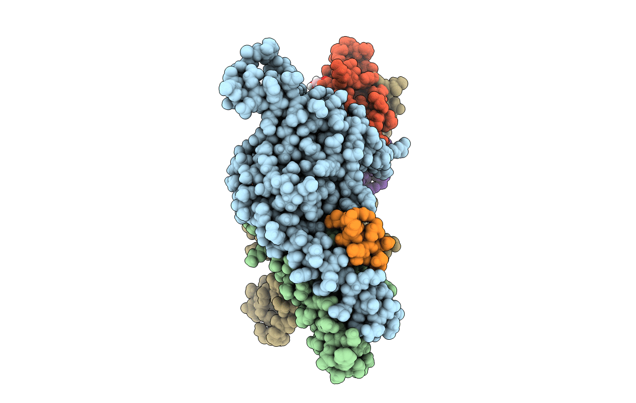
5OW5 image
Deposition Date 2017-08-30
Release Date 2018-07-11
Last Version Date 2024-01-17
Entry Detail
PDB ID:
5OW5
Keywords:
Title:
p60p80-CAMSAP complex
Biological Source:
Source Organism:
Mus musculus (Taxon ID: 10090)
Host Organism:
Method Details:
Experimental Method:
Resolution:
1.70 Å
R-Value Free:
0.23
R-Value Work:
0.19
R-Value Observed:
0.19
Space Group:
P 1 21 1
Macromolecular Entities
Structures with similar UniProt ID
Protein Blast
Polymer Type:polypeptide(L)
Molecule:Katanin p80 WD40 repeat-containing subunit B1
Gene (Uniprot):Katnb1
Chain IDs:A, C
Chain Length:212
Number of Molecules:2
Biological Source:Mus musculus
Structures with similar UniProt ID
Protein Blast
Polymer Type:polypeptide(L)
Molecule:Katanin p60 ATPase-containing subunit A1
Gene (Uniprot):Katna1
Chain IDs:B, D
Chain Length:80
Number of Molecules:2
Biological Source:Mus musculus
Structures with similar UniProt ID
Protein Blast
Polymer Type:polypeptide(L)
Molecule:Calmodulin-regulated spectrin-associated protein
Chain IDs:E, F
Chain Length:10
Number of Molecules:2
Biological Source:Mus musculus
Primary Citation
Structural Basis of Formation of the Microtubule Minus-End-Regulating CAMSAP-Katanin Complex.
Structure 26 375 382.e4 (2018)
PMID: 29395789 DOI: 10.1016/j.str.2017.12.017

Abstact

CAMSAP/Patronin family members regulate the organization and stability of microtubule minus ends in various systems ranging from mitotic spindles to differentiated epithelial cells and neurons. Mammalian CAMSAP2 and CAMSAP3 bind to growing microtubule minus ends, where they form stretches of stabilized microtubule lattice. The microtubule-severing ATPase katanin interacts with CAMSAPs and limits the length of CAMSAP-decorated microtubule stretches. Here, by using biochemical, biophysical, and structural approaches, we reveal that a short helical motif conserved in CAMSAP2 and CAMSAP3 binds to the heterodimer formed by the N- and C-terminal domains of katanin subunits p60 and p80, respectively. The identified CAMSAP-katanin binding mode is supported by mutational analysis and genome-editing experiments. It is strikingly similar to the one seen in the ASPM-katanin complex, which is responsible for microtubule minus-end regulation in mitotic spindles. Our work provides a general molecular mechanism for the cooperation of katanin with major microtubule minus-end regulators.

Legend

Protein

Chemical

Disease

Primary Citation of related structures
Feedback Form
Name
Email
Institute
Feedback