
Deposition Date
2017-08-23
Release Date
2018-09-05
Last Version Date
2024-01-17
Entry Detail
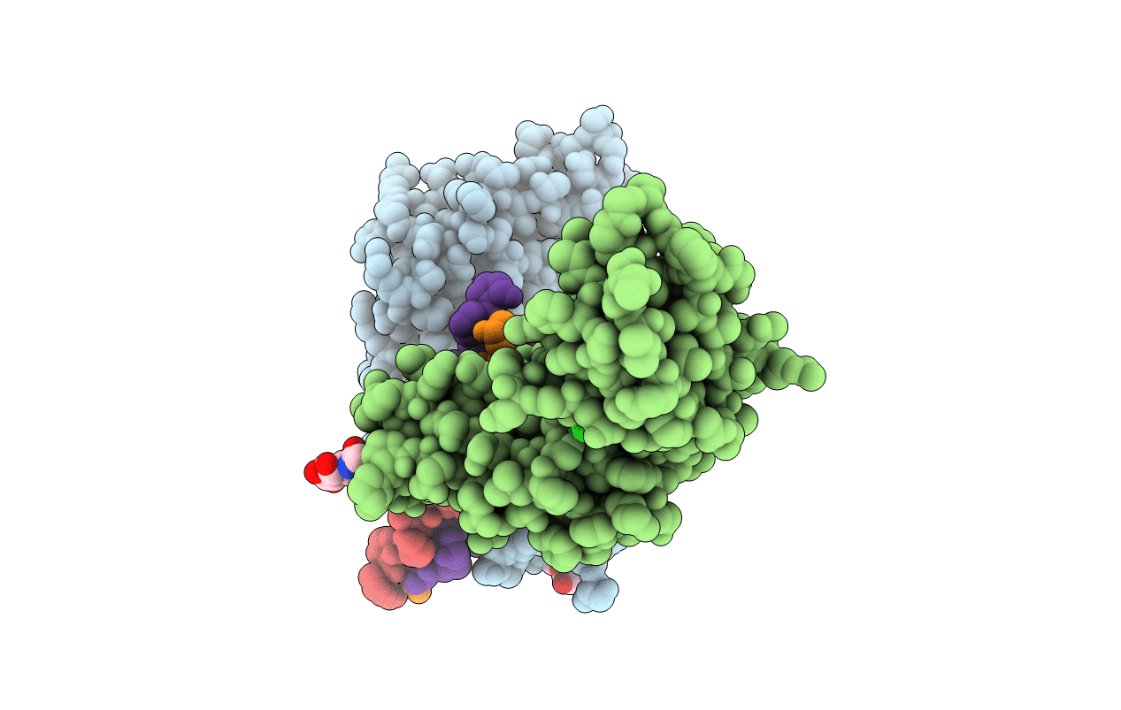
PDB ID:
5OU8
Keywords:
Title:
Crystal structure of Glycoprotein VI in complex with collagen-peptide (GPO)5
Biological Source:
Source Organism(s):
Homo sapiens (Taxon ID: 9606)
Expression System(s):
Method Details:
Experimental Method:
Resolution:
2.50 Å
R-Value Free:
0.25
R-Value Work:
0.20
R-Value Observed:
0.21
Space Group:
P 41 21 2
