
Deposition Date
2017-08-07
Release Date
2018-06-13
Last Version Date
2025-04-09
Entry Detail
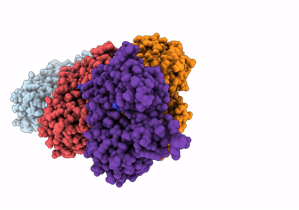
PDB ID:
5OOE
Keywords:
Title:
Cryo-EM structure of F-actin in complex with AppNHp (AMPPNP)
Biological Source:
Source Organism(s):
Oryctolagus cuniculus (Taxon ID: 9986)
Method Details:
Experimental Method:
Resolution:
3.60 Å
Aggregation State:
FILAMENT
Reconstruction Method:
SINGLE PARTICLE
