5ONJ image
Deposition Date 2017-08-03
Release Date 2018-08-29
Last Version Date 2024-11-13
Entry Detail
PDB ID:
5ONJ
Keywords:
Title:
YnDL in Complex with 5 amino acid (PGA) complex
Biological Source:
Source Organism:
Host Organism:
Method Details:
Experimental Method:
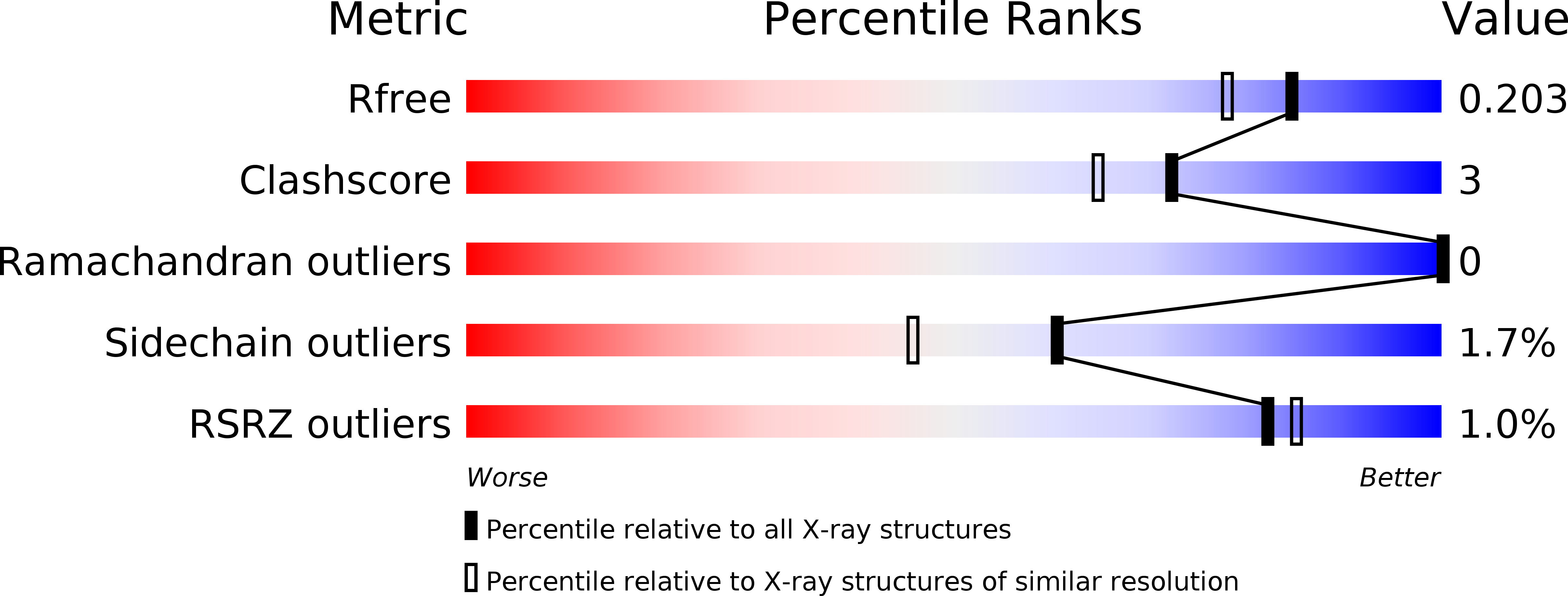
Resolution:
1.70 Å
R-Value Free:
0.20
R-Value Work:
0.16
R-Value Observed:
0.16
Space Group:
P 21 21 21
Macromolecular Entities
Polymer Type:polypeptide(L)
Molecule:YndL
Chain IDs:A
Chain Length:214
Number of Molecules:1
Biological Source:Bacillus subtilis
Polymer Type:polypeptide(L)
Molecule:(2R,3S,4R,5R,6R)-6-((1R,2R,3S,4R,6S)-4,6-DIAMINO-2,3-DIHYDROXYCYCLOHEXYLOXY)-5-AMINO-2-(AMINOMETHYL)-TETRAHYDRO-2H-PYRAN-3,4-DIOL
Chain IDs:B (auth: D)
Chain Length:6
Number of Molecules:1
Biological Source:synthetic construct
Ligand Molecules
Primary Citation
The structure of PghL hydrolase bound to its substrate poly-gamma-glutamate.
Febs J. 285 4575 4589 (2018)
PMID: 30387270 DOI: 10.1111/febs.14688

Abstact

The identification of new strategies to fight bacterial infections in view of the spread of multiple resistance to antibiotics has become mandatory. It has been demonstrated that several bacteria develop poly-γ-glutamic acid (γ-PGA) capsules as a protection from external insults and/or host defence systems. Among the pathogens that shield themselves in these capsules are Bacillus anthracis, Francisella tularensis and several Staphylococcus strains. These are important pathogens with a profound influence on human health. The recently characterised γ-PGA hydrolases, which can dismantle the γ-PGA-capsules, are an attractive new direction that can offer real hope for the development of alternatives to antibiotics, particularly in cases of multidrug resistant bacteria. We have characterised in detail the cleaving mechanism and stereospecificity of the enzyme PghL (previously named YndL) from Bacillus subtilis encoded by a gene of phagic origin and dramatically efficient in degrading the long polymeric chains of γ-PGA. We used X-ray crystallography to solve the three-dimensional structures of the enzyme in its zinc-free, zinc-bound and complexed forms. The protein crystallised with a γ-PGA hexapeptide substrate and thus reveals details of the interaction which could explain the stereospecificity observed and give hints on the catalytic mechanism of this class of hydrolytic enzymes.

Legend

Protein

Chemical

Disease

Primary Citation of related structures