
Deposition Date
2017-07-25
Release Date
2018-08-08
Last Version Date
2024-11-06
Entry Detail
PDB ID:
5OK3
Keywords:
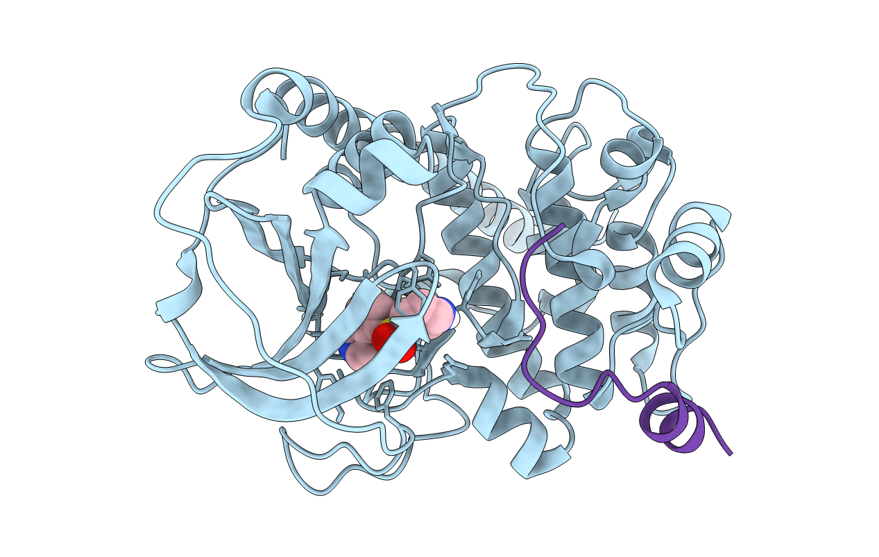
Title:
Crystal Structure of the Protein-Kinase A catalytic subunit from Criteculus Griseus in complex with compounds RKp241 and Fasudil
Biological Source:
Source Organism(s):
Cricetulus griseus (Taxon ID: 10029)
Expression System(s):
Method Details:
Experimental Method:
Resolution:
1.59 Å
R-Value Free:
0.17
R-Value Work:
0.13
R-Value Observed:
0.14
Space Group:
P 21 21 21
