5OHV image
Entry Detail
PDB ID:
5OHV
Title:
K33-specific affimer bound to K33 diUb
Biological Source:
Host Organism:
PDB Version:
Deposition Date:
2017-07-18
Release Date:
2017-10-04
Method Details:
Experimental Method:
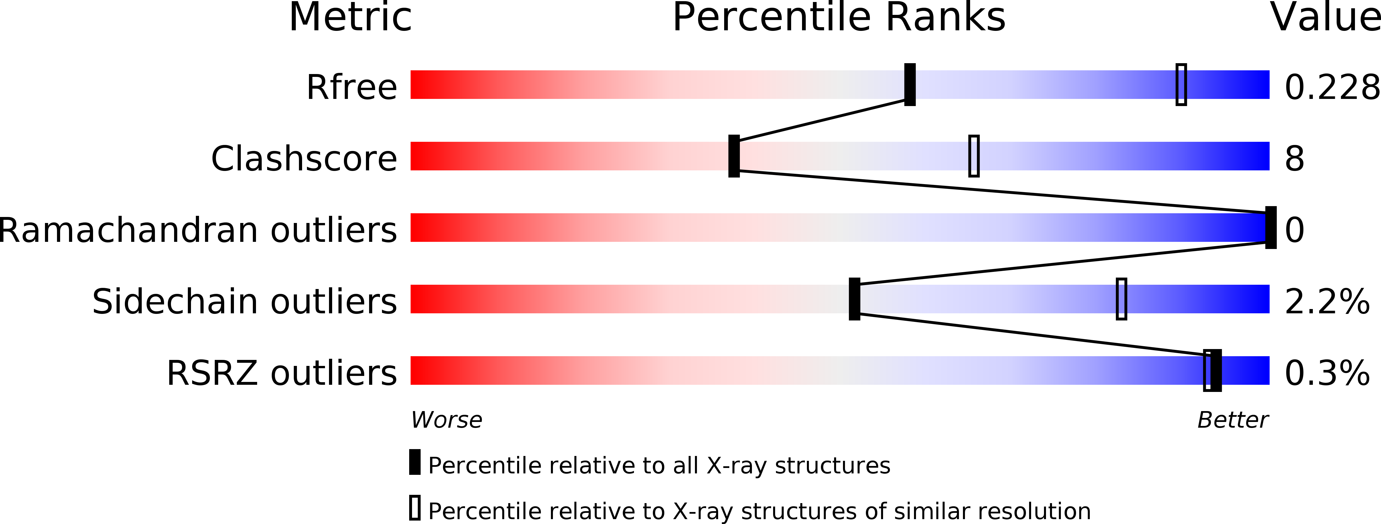
Resolution:
2.80 Å
R-Value Free:
0.22
R-Value Work:
0.17
R-Value Observed:
0.17
Space Group:
H 3
Macromolecular Entities
Polymer Type:polypeptide(L)
Description:Ubiquitin
Chain IDs:A, B (auth: C)
Chain Length:76
Number of Molecules:2
Biological Source:Homo sapiens
Polymer Type:polypeptide(L)
Description:K33-specific affimer
Chain IDs:C (auth: D), D (auth: B)
Chain Length:116
Number of Molecules:2
Biological Source:synthetic construct
Primary Citation
Ubiquitin Linkage-Specific Affimers Reveal Insights into K6-Linked Ubiquitin Signaling.
Mol. Cell 68 233 246.e5 (2017)
PMID: 28943312 DOI: 10.1016/j.molcel.2017.08.020

Abstact

Several ubiquitin chain types have remained unstudied, mainly because tools and techniques to detect these posttranslational modifications are scarce. Linkage-specific antibodies have shaped our understanding of the roles and dynamics of polyubiquitin signals but are available for only five out of eight linkage types. We here characterize K6- and K33-linkage-specific "affimer" reagents as high-affinity ubiquitin interactors. Crystal structures of affimers bound to their cognate chain types reveal mechanisms of specificity and a K11 cross-reactivity in the K33 affimer. Structure-guided improvements yield superior affinity reagents suitable for western blotting, confocal fluorescence microscopy and pull-down applications. This allowed us to identify RNF144A and RNF144B as E3 ligases that assemble K6-, K11-, and K48-linked polyubiquitin in vitro. A protocol to enrich K6-ubiquitinated proteins from cells identifies HUWE1 as a main E3 ligase for this chain type, and we show that mitofusin-2 is modified with K6-linked polyubiquitin in a HUWE1-dependent manner.

Legend

Protein

Chemical

Disease

Primary Citation of related structures