5OH0 image
Deposition Date 2017-07-13
Release Date 2017-11-22
Last Version Date 2024-11-20
Entry Detail
PDB ID:
5OH0
Keywords:
Title:
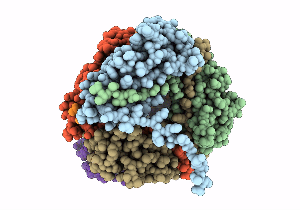
The Cryo-Electron Microscopy Structure of the Type 1 Chaperone-Usher Pilus Rod
Biological Source:
Source Organism:
Host Organism:
Method Details:
Experimental Method:
Resolution:
4.20 Å
Aggregation State:
HELICAL ARRAY
Reconstruction Method:
HELICAL
Macromolecular Entities
Polymer Type:polypeptide(L)
Molecule:Type-1 fimbrial protein, A chain
Gene (Uniprot):fimA
Chain IDs:A, B, C, D, E, F
Chain Length:159
Number of Molecules:6
Biological Source:Escherichia coli J96
Ligand Molecules
Primary Citation
The Cryoelectron Microscopy Structure of the Type 1 Chaperone-Usher Pilus Rod.
Structure 25 1829 1838.e4 (2017)
PMID: 29129382 DOI: 10.1016/j.str.2017.10.004

Abstact

Adhesive chaperone-usher pili are long, supramolecular protein fibers displayed on the surface of many bacterial pathogens. The type 1 and P pili of uropathogenic Escherichia coli (UPEC) play important roles during urinary tract colonization, mediating attachment to the bladder and kidney, respectively. The biomechanical properties of the helical pilus rods allow them to reversibly uncoil in response to flow-induced forces, allowing UPEC to retain a foothold in the unique and hostile environment of the urinary tract. Here we provide the 4.2-Å resolution cryo-EM structure of the type 1 pilus rod, which together with the previous P pilus rod structure rationalizes the remarkable "spring-like" properties of chaperone-usher pili. The cryo-EM structure of the type 1 pilus rod differs in its helical parameters from the structure determined previously by a hybrid approach. We provide evidence that these structural differences originate from different quaternary structures of pili assembled in vivo and in vitro.

Legend

Protein

Chemical

Disease

Primary Citation of related structures