5OGH image
Deposition Date 2017-07-13
Release Date 2018-08-01
Last Version Date 2024-11-13
Entry Detail
PDB ID:
5OGH
Keywords:
Title:
Structure of RNase A at high resolution (1.16 A) in complex with 3'-CMP and sulphate ions
Biological Source:
Source Organism:
Bos taurus (Taxon ID: 9913)
Method Details:
Experimental Method:
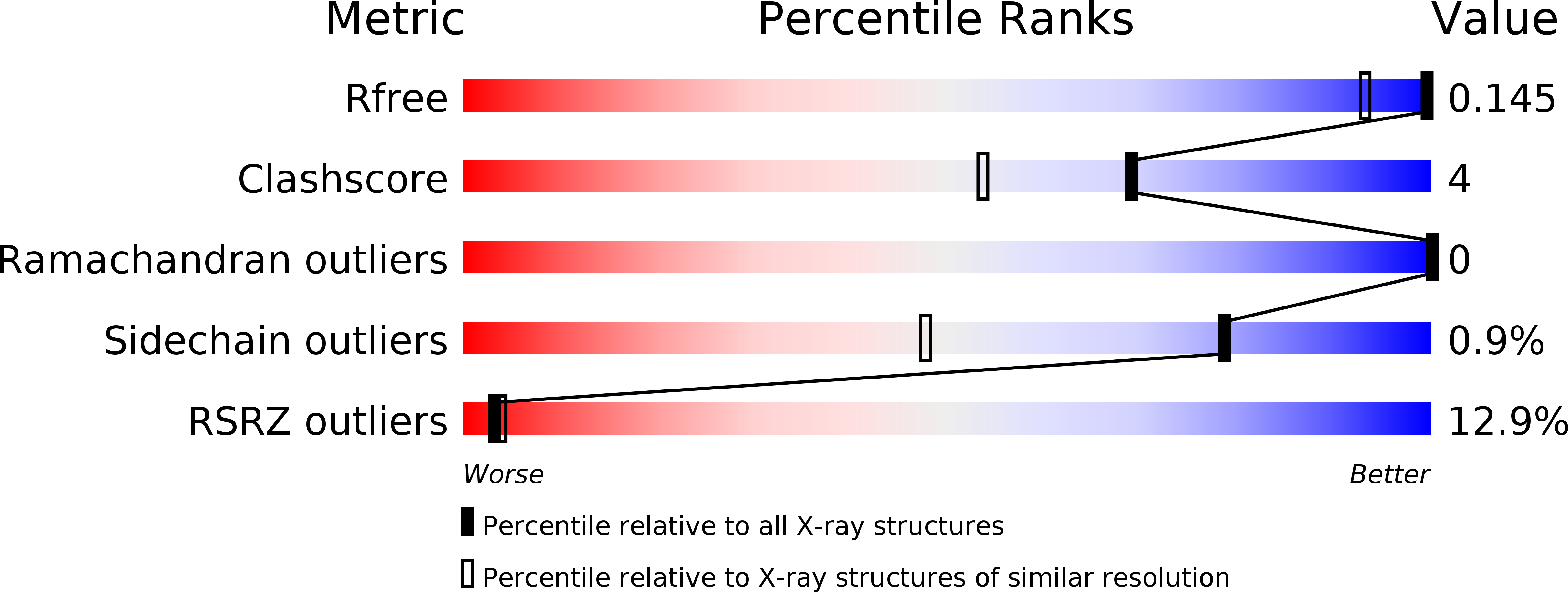
Resolution:
1.16 Å
R-Value Free:
0.13
R-Value Work:
0.12
R-Value Observed:
0.12
Space Group:
P 32 2 1
Macromolecular Entities
Polymer Type:polypeptide(L)
Molecule:Ribonuclease pancreatic
Gene (Uniprot):RNASE1
Chain IDs:A
Chain Length:124
Number of Molecules:1
Biological Source:Bos taurus
Primary Citation
Characterization of an RNase with two catalytic centers. Human RNase6 catalytic and phosphate-binding site arrangement favors the endonuclease cleavage of polymeric substrates.
Biochim Biophys Acta Gen Subj 1863 105 117 (2019)
PMID: 30287244 DOI: 10.1016/j.bbagen.2018.09.021

Abstact

BACKGROUND Human RNase6 is a small cationic antimicrobial protein that belongs to the vertebrate RNaseA superfamily. All members share a common catalytic mechanism, which involves a conserved catalytic triad, constituted by two histidines and a lysine (His15/His122/Lys38 in RNase6 corresponding to His12/His119/Lys41 in RNaseA). Recently, our first crystal structure of human RNase6 identified an additional His pair (His36/His39) and suggested the presence of a secondary active site. METHODS In this work we have explored RNase6 and RNaseA subsite architecture by X-ray crystallography, site-directed mutagenesis and kinetic characterization. RESULTS The analysis of two novel crystal structures of RNase6 in complex with phosphate anions at atomic resolution locates a total of nine binding sites and reveals the contribution of Lys87 to phosphate-binding at the secondary active center. Contribution of the second catalytic triad residues to the enzyme activity is confirmed by mutagenesis. RNase6 catalytic site architecture has been compared with an RNaseA engineered variant where a phosphate-binding subsite is converted into a secondary catalytic center (RNaseA-K7H/R10H). CONCLUSIONS We have identified the residues that participate in RNase6 second catalytic triad (His36/His39/Lys87) and secondary phosphate-binding sites. To note, residues His39 and Lys87 are unique within higher primates. The RNaseA/RNase6 side-by-side comparison correlates the presence of a dual active site in RNase6 with a favored endonuclease-type cleavage pattern. GENERAL SIGNIFICANCE An RNase dual catalytic and extended binding site arrangement facilitates the cleavage of polymeric substrates. This is the first report of the presence of two catalytic centers in a single monomer within the RNaseA superfamily.

Legend

Protein

Chemical

Disease

Primary Citation of related structures