Deposition Date
2017-07-12
Release Date
2018-10-24
Last Version Date
2024-01-17
Entry Detail
PDB ID:
5OGB
Keywords:
Title:
Human Cellular Retinoic Acid Binding Protein II (CRABPII) with bound synthetic retinoid DC360.
Biological Source:
Source Organism(s):
Homo sapiens (Taxon ID: 9606)
Expression System(s):
Method Details:
Experimental Method:
Resolution:
1.80 Å
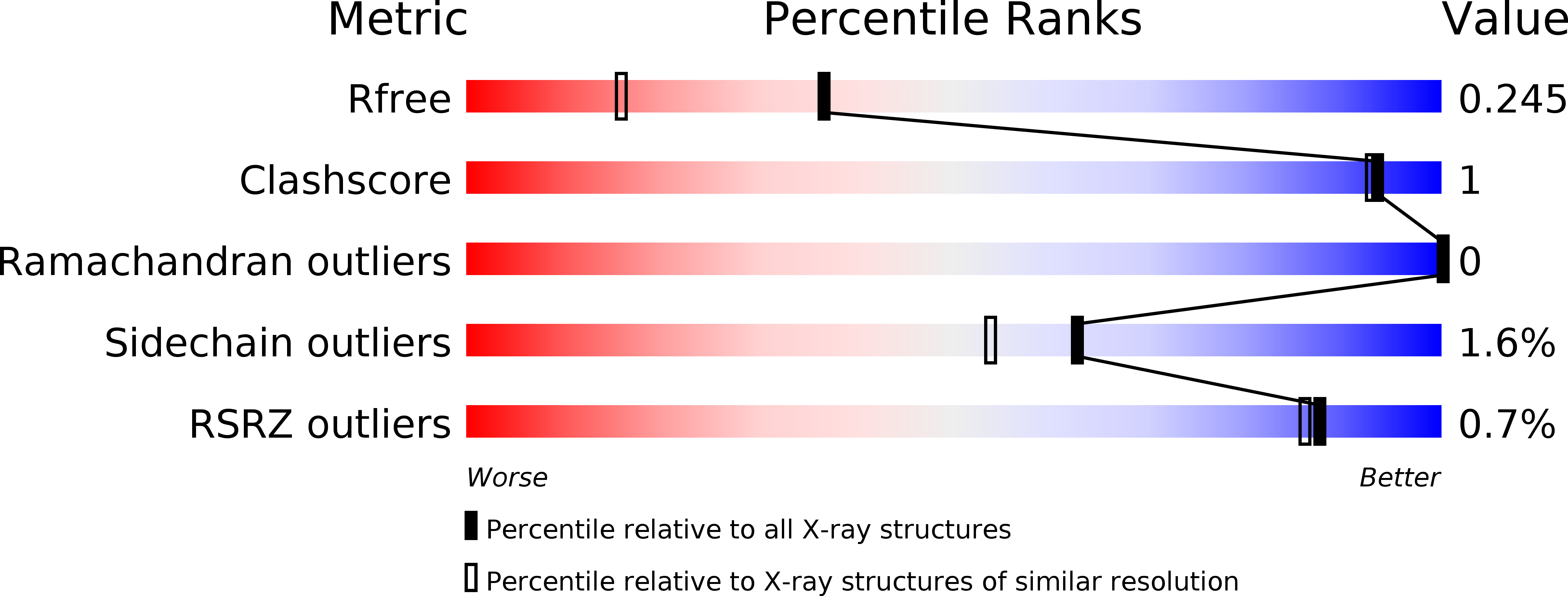
R-Value Free:
0.23
R-Value Work:
0.17
R-Value Observed:
0.17
Space Group:
P 1 21 1