Deposition Date
2017-07-11
Release Date
2017-09-27
Last Version Date
2024-01-17
Entry Detail
PDB ID:
5OFQ
Keywords:
Title:
Crystal structure of substrate-free CYP109A2 from Bacillus megaterium
Biological Source:
Source Organism(s):
Bacillus megaterium (strain DSM 319) (Taxon ID: 592022)
Expression System(s):
Method Details:
Experimental Method:
Resolution:
2.70 Å
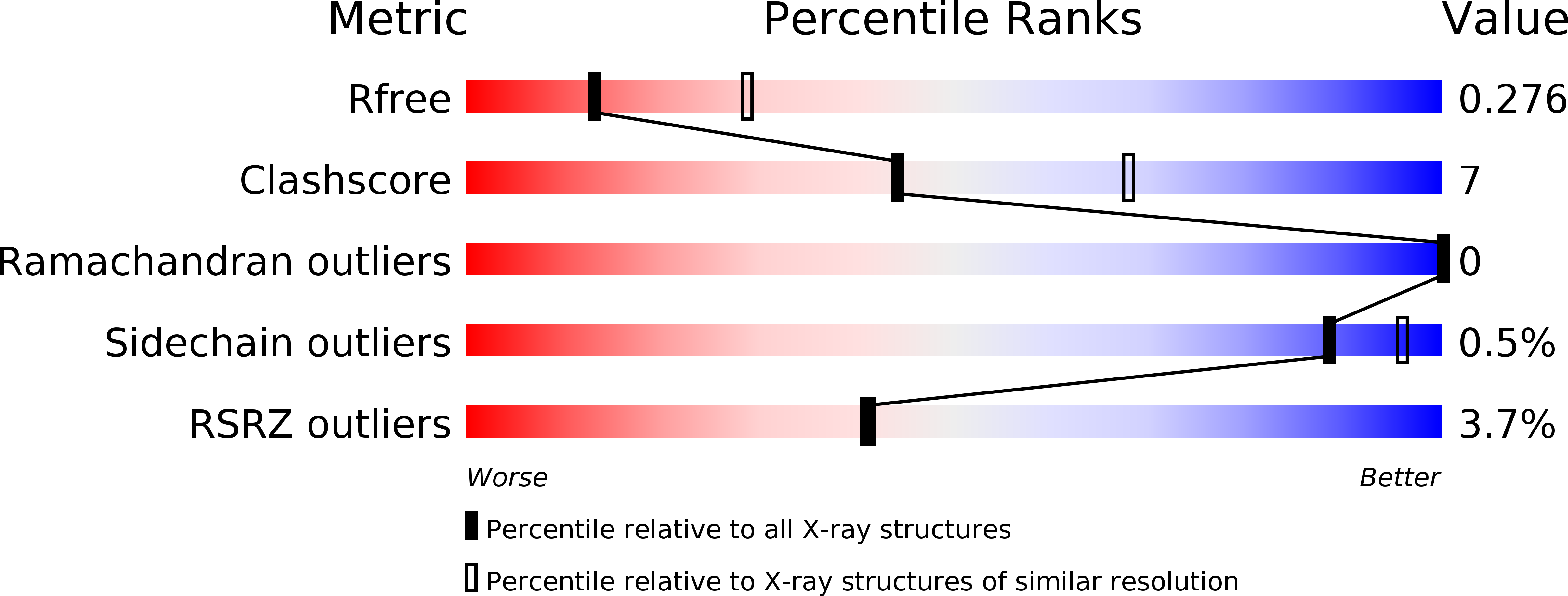
R-Value Free:
0.27
R-Value Work:
0.22
R-Value Observed:
0.22
Space Group:
P 21 21 21