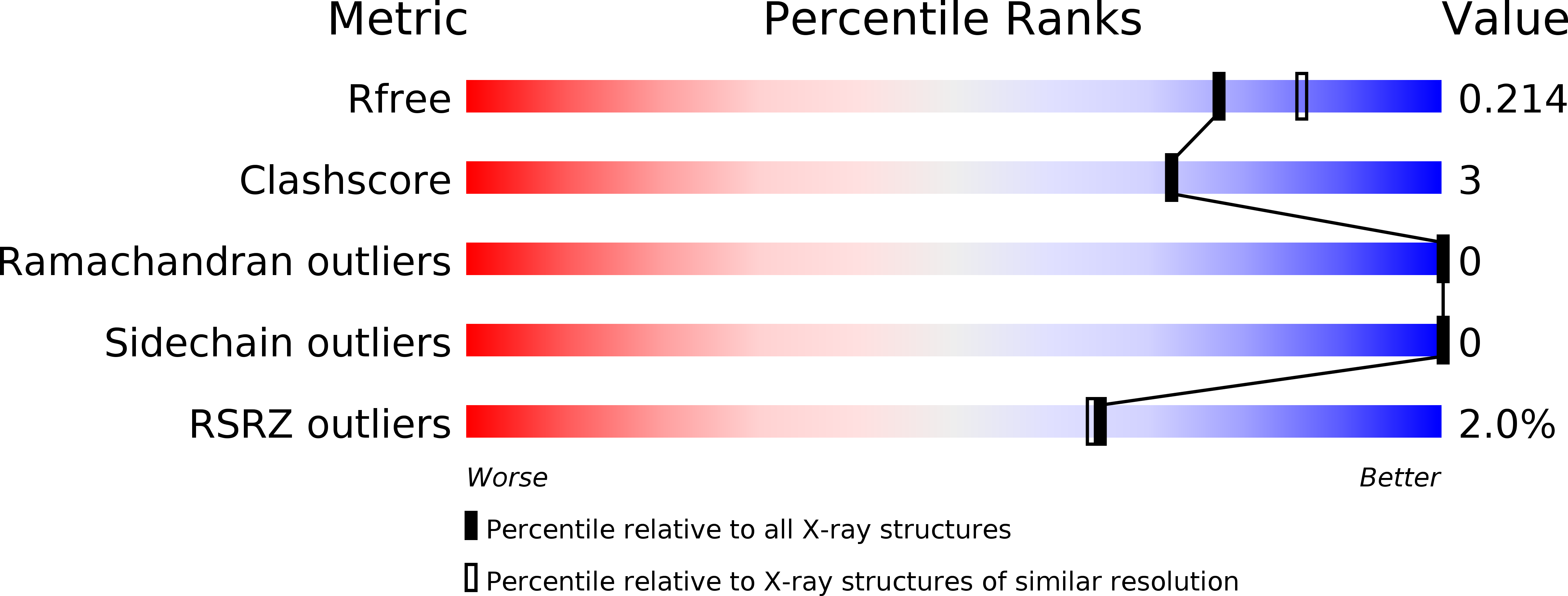
5ODT image
Entry Detail
PDB ID:
5ODT
Keywords:
Title:
Aurora-A in complex with TACC3
Biological Source:
Source Organism:
PDB Version:
Deposition Date:
2017-07-06
Release Date:
2018-03-14
Method Details:
Experimental Method:
Resolution:
2.02 Å
R-Value Free:
0.21
R-Value Work:
0.17
R-Value Observed:
0.17
Space Group:
I 2 3
Macromolecular Entities
Polymer Type:polypeptide(L)
Description:Aurora kinase A
Mutations:D274N, C290A, C393A
Chain IDs:A
Chain Length:283
Number of Molecules:1
Biological Source:Homo sapiens
Polymer Type:polypeptide(L)
Description:Transforming acidic coiled-coil-containing protein 3
Chain IDs:B
Chain Length:46
Number of Molecules:1
Biological Source:Homo sapiens
Primary Citation
Mitotic spindle association of TACC3 requires Aurora-A-dependent stabilization of a cryptic alpha-helix.
EMBO J. 37 ? ? (2018)
PMID: 29510984 DOI: 10.15252/embj.201797902

Abstact

Aurora-A regulates the recruitment of TACC3 to the mitotic spindle through a phospho-dependent interaction with clathrin heavy chain (CHC). Here, we describe the structural basis of these interactions, mediated by three motifs in a disordered region of TACC3. A hydrophobic docking motif binds to a previously uncharacterized pocket on Aurora-A that is blocked in most kinases. Abrogation of the docking motif causes a delay in late mitosis, consistent with the cellular distribution of Aurora-A complexes. Phosphorylation of Ser558 engages a conformational switch in a second motif from a disordered state, needed to bind the kinase active site, into a helical conformation. The helix extends into a third, adjacent motif that is recognized by a helical-repeat region of CHC, not a recognized phospho-reader domain. This potentially widespread mechanism of phospho-recognition provides greater flexibility to tune the molecular details of the interaction than canonical recognition motifs that are dominated by phosphate binding.

Legend

Protein

Chemical

Disease

Primary Citation of related structures