Deposition Date
2017-07-03
Release Date
2018-03-28
Last Version Date
2024-11-13
Entry Detail
PDB ID:
5OCV
Keywords:
Title:
A Rare Lysozyme Crystal Form Solved Using High-Redundancy 3D Electron Diffraction Data from Micron-Sized Needle Shaped Crystals
Biological Source:
Source Organism(s):
Gallus gallus (Taxon ID: 9031)
Method Details:
Experimental Method:
Resolution:
2.20 Å
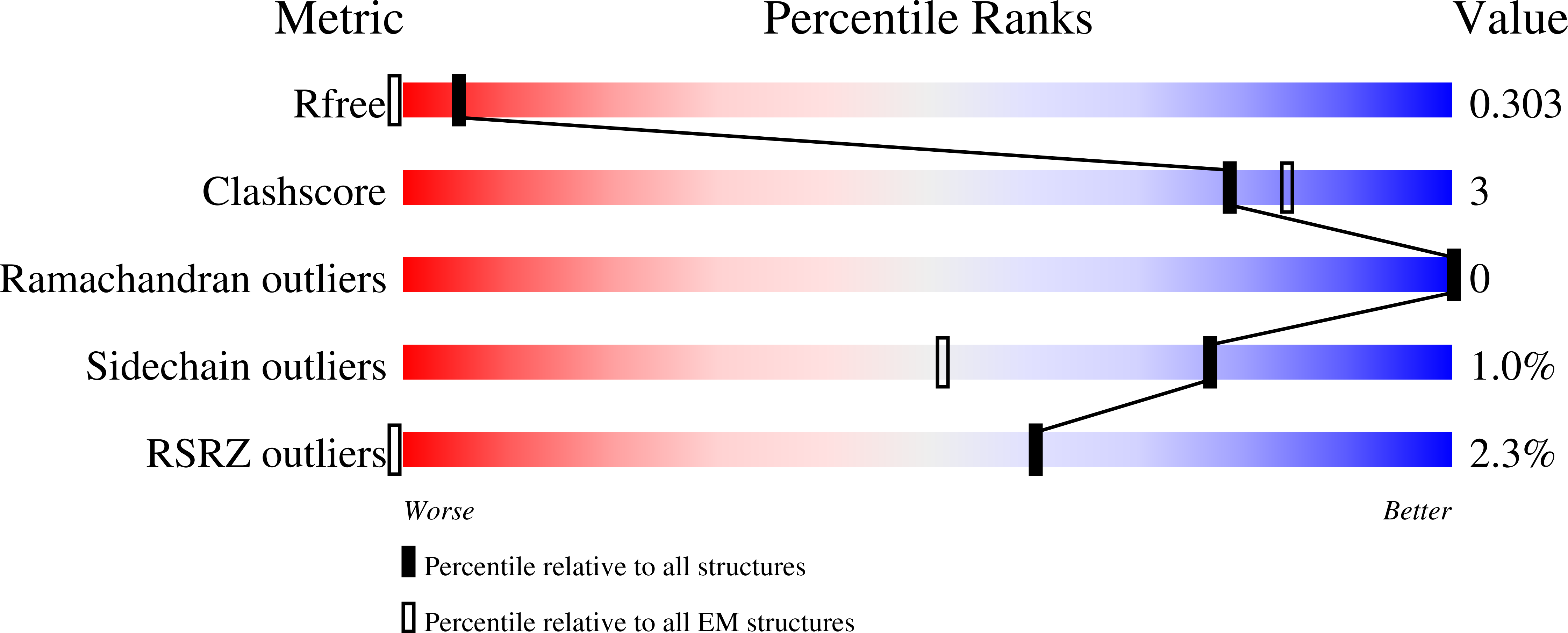
R-Value Free:
0.27
R-Value Work:
0.23
R-Value Observed:
0.23
Space Group:
P 21 21 2