5OCC image
Deposition Date 2017-06-30
Release Date 2018-07-25
Last Version Date 2024-11-06
Entry Detail
PDB ID:
5OCC
Keywords:
Title:
Crystal structure of CD32b (Fc Gamma Receptor IIb) in complex with Human IgG1 Fab fragment (6G08)
Biological Source:
Source Organism:
Homo sapiens (Taxon ID: 9606)
Host Organism:
Method Details:
Experimental Method:
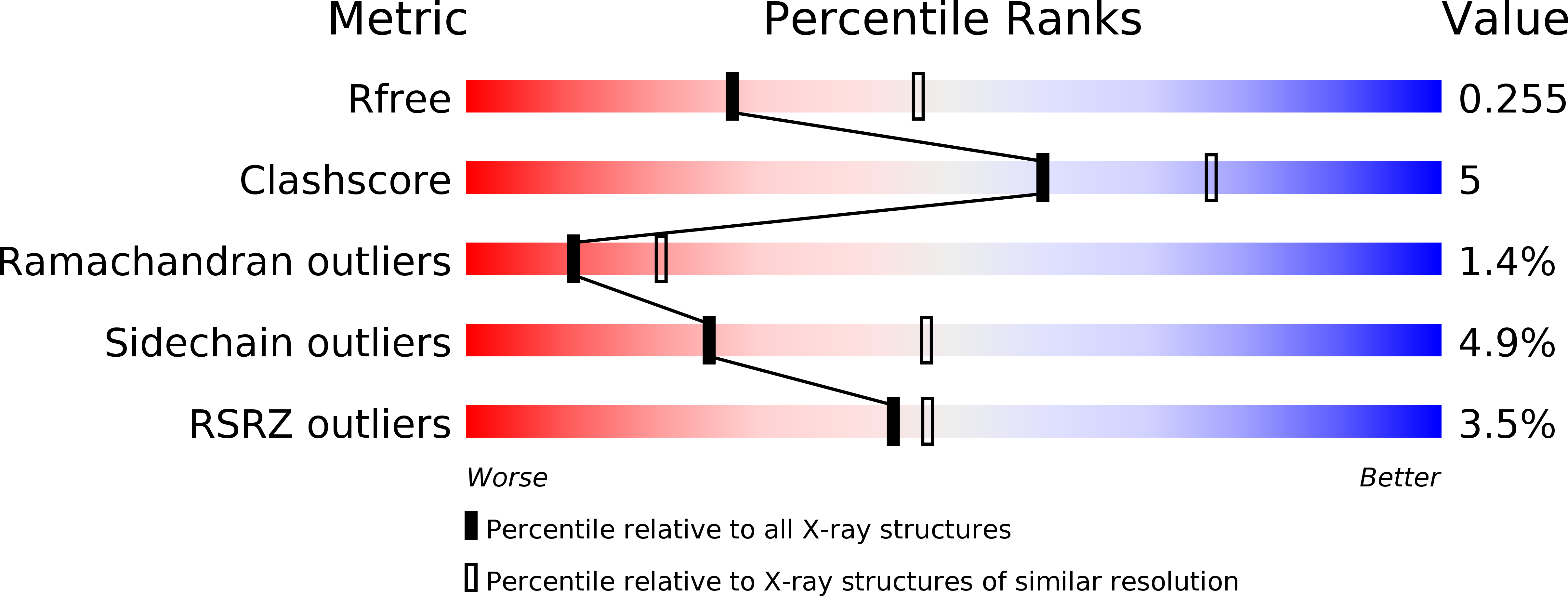
Resolution:
2.50 Å
R-Value Free:
0.26
R-Value Work:
0.19
R-Value Observed:
0.19
Space Group:
P 21 21 21
Macromolecular Entities
Polymer Type:polypeptide(L)
Molecule:Low affinity immunoglobulin gamma Fc region receptor II-b
Gene (Uniprot):FCGR2B
Chain IDs:A
Chain Length:176
Number of Molecules:1
Biological Source:Homo sapiens
Polymer Type:polypeptide(L)
Molecule:6G08 Fab heavy chain
Chain IDs:B (auth: H)
Chain Length:222
Number of Molecules:1
Biological Source:Homo sapiens
Polymer Type:polypeptide(L)
Molecule:6G08 Fab Light Chain
Chain IDs:C (auth: L)
Chain Length:217
Number of Molecules:1
Biological Source:Homo sapiens
Primary Citation
Evaluating Anti-CD32b F(ab) Conformation Using Molecular Dynamics and Small-Angle X-Ray Scattering.
Biophys. J. 115 289 299 (2018)
PMID: 30021105 DOI: 10.1016/j.bpj.2018.03.040

Abstact

Complementary strategies of small-angle x-ray scattering (SAXS) and crystallographic analysis are often used to determine atomistic three-dimensional models of macromolecules and their variability in solution. This combination of techniques is particularly valuable when applied to macromolecular complexes to detect changes within the individual binding partners. Here, we determine the x-ray crystallographic structure of a F(ab) fragment in complex with CD32b, the only inhibitory Fc-γ receptor in humans, and compare the structure of the F(ab) from the crystal complex to SAXS data for the F(ab) alone in solution. We investigate changes in F(ab) structure by predicting theoretical scattering profiles for atomistic structures extracted from molecular dynamics (MD) simulations of the F(ab) and assessing the agreement of these structures to our experimental SAXS data. Through principal component analysis, we are able to extract principal motions observed during the MD trajectory and evaluate the influence of these motions on the agreement of structures to the F(ab) SAXS data. Changes in the F(ab) elbow angle were found to be important to reach agreement with the experimental data; however, further discrepancies were apparent between our F(ab) structure from the crystal complex and SAXS data. By analyzing multiple MD structures observed in similar regions of the principal component analysis, we were able to pinpoint these discrepancies to a specific loop region in the F(ab) heavy chain. This method, therefore, not only allows determination of global changes but also allows identification of localized motions important for determining the agreement between atomistic structures and SAXS data. In this particular case, the findings allowed us to discount the hypothesis that structural changes were induced upon complex formation, a significant find informing the drug development process. The methodology described here is generally applicable to deconvolute global and local changes of macromolecular structures and is well suited to other systems.

Legend

Protein

Chemical

Disease

Primary Citation of related structures