Deposition Date
2017-06-26
Release Date
2018-07-25
Last Version Date
2024-01-17
Entry Detail
PDB ID:
5OBB
Keywords:
Title:
Structure of a modified mouse H chain ferritin with a lanthanide binding motif in complex with Terbium
Biological Source:
Source Organism(s):
Mus musculus (Taxon ID: 10090)
Expression System(s):
Method Details:
Experimental Method:
Resolution:
2.65 Å
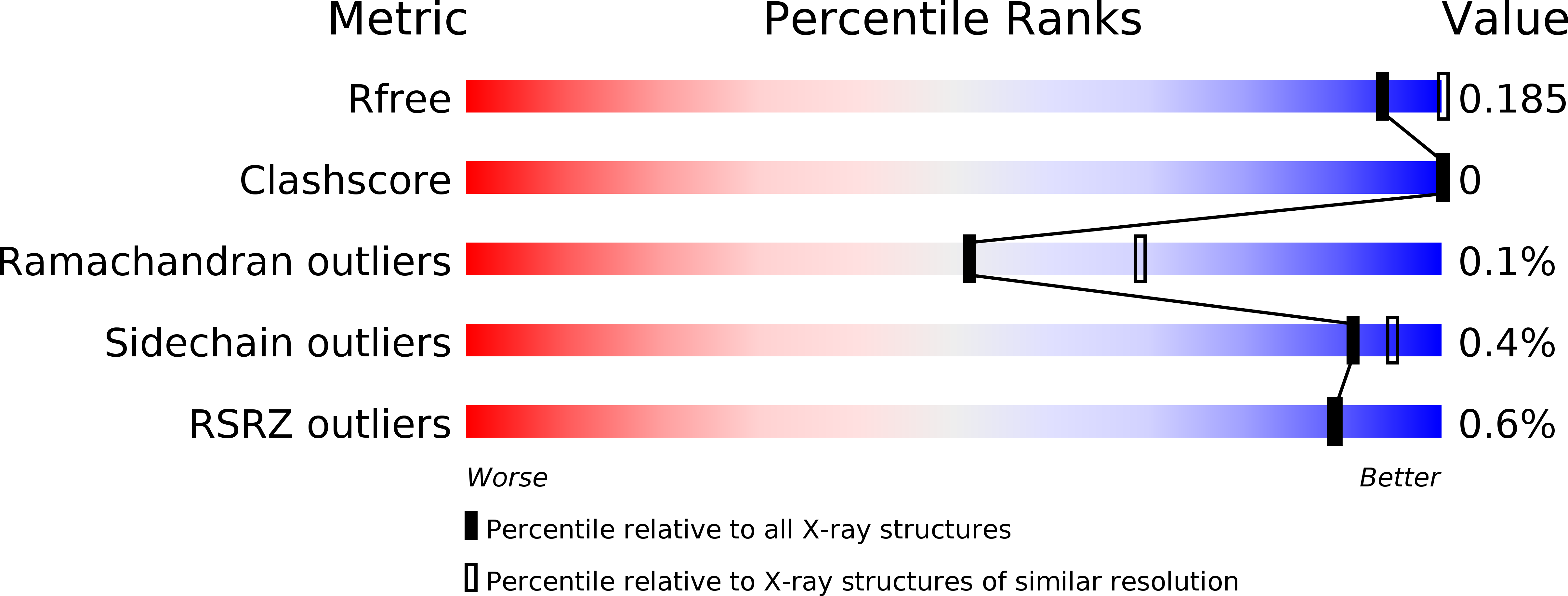
R-Value Free:
0.17
R-Value Work:
0.16
R-Value Observed:
0.16
Space Group:
I 2 2 2